中国组织工程研究 ›› 2022, Vol. 26 ›› Issue (22): 3550-3555.doi: 10.12307/2022.304

• 复合支架材料 composite scaffold materials • 上一篇    下一篇

基于低温3D打印丝素蛋白/Ⅰ型胶原/羟基磷灰石支架的力学性能

孟露露1,2,刘  浩3,刘  涵3,4,张  军3,李瑞欣3,高丽兰1,2   

  1. 1天津理工大学,天津市先进机电系统设计与智能控制重点实验室,天津市  300384;2机电工程国家级实验教学示范中心(天津理工大学),天津市  300384;3南开大学附属口腔医院,天津市口腔医院,天津市口腔功能重建重点实验室,天津市  300041;4南开大学,天津市  300071
  • 收稿日期:2021-01-28 修回日期:2021-03-12 接受日期:2021-06-09 出版日期:2022-08-08 发布日期:2022-01-12
  • 通讯作者: 李瑞欣,副研究员,博士,南开大学附属口腔医院,天津市口腔医院,天津市口腔功能重建重点实验室,天津市 300041 高丽兰,教授,博士,天津理工大学,天津市先进机电系统设计与智能控制重点实验室,天津市 300384;机电工程国家级实验教学示范中心(天津理工大学),天津市 300384
  • 作者简介:孟露露,男,1992年生,山西省怀仁市人,汉族,天津理工大学在读硕士,主要从事生物力学研究。
  • 基金资助:
    国家自然科学基金项目 (11972198),项目负责人,李瑞欣;天津市临床医学重点学科专项 (HWZX001),项目负责人:刘浩;天津市重大疾病防治科技重大专项(19ZXDBSY00070),项目负责人:刘浩

Mechanical properties of silk fibroin/type I collagen/hydroxyapatite scaffolds based on low-temperature 3D printing

Meng Lulu1, 2, Liu Hao3, Liu Han3, 4, Zhang Jun3, Li Ruixin3, Gao Lilan1, 2   

  1. 1Tianjin Key Laboratory for Advanced Mechatronic System Design and Intelligent Control, School of Mechanical Engineering, Tianjin University of Technology; 2National Demonstration Center for Experimental Mechanical and Electrical Engineering Education (Tianjin University of Technology); 3the Affiliated Stomatological Hospital of Nankai University, Tianjin Stomatological Hospital, Tianjin Key Laboratory of Oral and Maxillofacial Function Reconstruction; 4Nankai University
  • Received:2021-01-28 Revised:2021-03-12 Accepted:2021-06-09 Online:2022-08-08 Published:2022-01-12
  • Contact: Li Ruixin, Associate researcher, MD, the Affiliated Stomatological Hospital of Nankai University, Tianjin Stomatological Hospital, Tianjin Key Laboratory of Oral and Maxillofacial Function Reconstruction, Tianjin 300041, China Gao Lilan, Professor, MD, Tianjin Key Laboratory for Advanced Mechatronic System Design and Intelligent Control, School of Mechanical Engineering, Tianjin University of Technology, Tianjin 300384, China; National Demonstration Center for Experimental Mechanical and Electrical Engineering Education (Tianjin University of Technology), Tianjin 300384, China
  • About author:Meng Lulu, Master candidate, Tianjin Key Laboratory for Advanced Mechatronic System Design and Intelligent Control, School of Mechanical Engineering, Tianjin University of Technology, Tianjin 300384, China; National Demonstration Center for Experimental Mechanical and Electrical Engineering Education (Tianjin University of Technology), Tianjin 300384, China
  • Supported by:
    the National Natural Science Foundation of China, No. 11972198 (to LRX); the Key Discipline of Clinical Medicine of Tianjin, No. HWZX001 (to LH); Major Disease Prevention and Control Technology Project of Tianjin, No. 19ZXDBSY00070 (to LH)

摘要:

文题释义:
应力松弛:在总应变不变的条件下,由于黏弹性材料内部的黏性应变分量随时间不断增长,使回弹性应变分量随时间逐渐降低,从而导致变形恢复力随时间逐渐降低的现象。
蠕变:蠕变实验方法有很多种类,如通过对材料试件施加恒定的压力以研究材料压缩蠕变性能,还可以对材料施加恒定扭力以研究材料的扭转蠕变性能等。

背景:随着3D打印技术在组织工程的快速发展,通过3D打印制备出多种支架材料被广泛用于下颌骨缺损修复,3D打印技术为下颌骨缺损修复带来了新的可能。
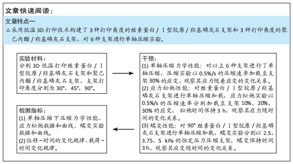
目的:采用低温3D打印技术构建三维仿生骨支架材料,精确控制支架材料内部结构,并对其进行力学性能分析。
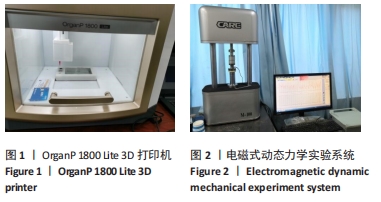
方法:在同等体积下,通过改变打印支架线束与线束的交错角度,采用低温3D打印技术分别打印不同角度(30°,45°,90°)的丝素蛋白/Ⅰ型胶原/羟基磷灰石支架与聚己内酯/羟基磷灰石支架(共6组支架)。单轴压缩力学实验以0.5%/s的压缩速率加载6组支架,压缩至30%的应变,观察应力随着应变的变化关系;应力松弛实验以0.5%/s的压缩速率分别加载至3种打印角度丝素蛋白/Ⅰ型胶原/羟基磷灰石支架10%,20%,30%的应变,松弛保持时间3 h,观察应力随时间的变化关系;蠕变实验分别以2.5,3.75,5 kPa的恒定压力压缩打印角度为90°的丝素蛋白/Ⅰ型胶原/羟基磷灰石支架,蠕变保持时间3 h,观察应变随时间的变化关系。
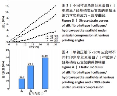
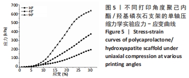
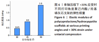
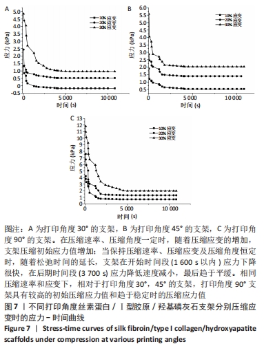
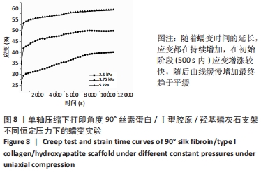
结果与结论:①单轴压缩力学实验:对于丝素蛋白/Ⅰ型胶原/羟基磷灰石支架和聚己内酯/羟基磷灰石支架,相同压缩应变下打印角度为90°支架的杨氏模量高于打印角度为30°,45°的支架。②应力松弛实验:当保持压缩速率、压缩应变及压缩角度恒定时,丝素蛋白/Ⅰ型胶原/羟基磷灰石支架的应力随松弛时间的延伸先快速降低,然后缓慢降低,随着松弛时间的延长,支架在开始时间段(1 600 s以内)应力下降很快,在后期时间段(3 700 s)应力降低速度减小,最后趋于平缓;当保持压缩速率、压缩应变恒定时,相对于打印角度为30°,45°的支架,打印角度为90°丝素蛋白/Ⅰ型胶原/羟基磷灰石支架的应力初值及趋于平稳时的应力值较高;当压缩速率及压缩角度恒定时,随着压缩应变值增加,丝素蛋白/Ⅰ型胶原/羟基磷灰石支架的应力值增加。③蠕变实验:随着蠕变时间的延长,90°的丝素蛋白/Ⅰ型胶原/羟基磷灰石支架的应变增加,在初始阶段(500 s内)应变增涨较快,随后应变缓慢增加最终趋于平缓,2.5 kPa下支架的应变变化范围为35%到55%,3.75 kPa下支架的应变变化范围从43%到57%,5 kPa下支架的应变变化范围从45%到57%。

https://orcid.org/0000-0003-3758-0910 (孟露露) ;https://orcid.org/0000-0002-7288-6686 (高丽兰)

中国组织工程研究杂志出版内容重点:生物材料;骨生物材料口腔生物材料纳米材料缓释材料材料相容性;组织工程

关键词: 3D打印, 生物支架, 丝素蛋白, Ⅰ型胶原, 羟基磷灰石, 单轴压缩, 生物力学

Abstract: BACKGROUND: With the rapid development of 3D printing technology in tissue engineering, a variety of scaffold materials prepared by 3D printing are widely used in mandibular defect repair. 3D printing technology brings new possibilities for mandibular defect repair. 
OBJECTIVE: The 3D bionic bone scaffold material was constructed by low-temperature 3D printing technology, and the internal structure of the scaffold was precisely controlled, and the mechanical properties of the scaffold were analyzed.
METHODS: Under the same volume, by changing the interlacing angle of the printed scaffold wire harness and the wire harness, low-temperature 3D printing technology was used to print silk fibroin/type I collagen/hydroxyapatite scaffolds with different angles (30°, 45°, 90°) and polycaprolactone/hydroxyapatite scaffolds (a total of 6 sets of scaffolds). The uniaxial compression mechanics experiment was loaded to 6 groups of scaffolds at a compression rate of 0.5%/s, compressed to 30% strain to observe the relationship between stress and strain. The stress relaxation experiment was applied to the three printing angles of silk fibroin/collagen I/hydroxyapatite scaffolds at a compression rate of 0.5%/s at 10%, 20%, and 30% strain, and the relaxation retention time was 3 hours to observe the relationship between stress and time. In the creep experiment, the silk fibroin/collagen I/hydroxyapatite scaffold with an angle of 90° was compressed and printed at a constant pressure of 2.5, 3.75, and 5 kPa, and the creep retention time was 3 hours to observe the relationship between strain and time.
RESULTS AND CONCLUSION: (1) Uniaxial compression mechanics experiment: The mechanical properties of 3D printed silk fibroin/type I collagen/hydroxyapatite scaffold and polycaprolactone/hydroxyapatite scaffold compression showed that the 90° scaffold had higher Young’s modulus than the 30° and 45° scaffolds under the same compression strain. (2) Stress relaxation experiment: When the compression rate, compression strain and compression angle were constant, the stress of silk fibroin/type I collagen/hydroxyapatite scaffold decreased rapidly with the extension of relaxation time, then slowly decreased. With the extension of relaxation time, the stress of the scaffold decreased rapidly in the beginning time (within 1 600 seconds), and in the later period (3 700 seconds). When the compression rate and compression strain were kept constant, the initial and stable stress values of silk fibroin/type I collagen/hydroxyapatite scaffolds with 90° printing angle were higher than those of scaffolds with 30° and 45° printing angle. When the compression rate and compression angle were constant, the initial and stable stress values of silk fibroin/type I collagen/hydroxyapatite scaffolds increased with the increase of compression strain. (3) Creep test: With the extension of creep time, the strain of 90° silk fibroin/type I collagen / hydroxyapatite scaffolds increased rapidly in the initial stage (within 500 seconds), then increased slowly, and finally leveled off. The strain range of the scaffolds was 35% to 55% at 2.5 kPa, and 43% to 57% at 3.75 kPa. The strain of the scaffold ranged from 45% to 57% under 5 kPa. 

Key words: 3D printing, biological scaffold, silk fibroin, type I collagen, hydroxyapatite, uniaxial compression, biomechanics

中图分类号: