中国组织工程研究 ›› 2021, Vol. 25 ›› Issue (34): 5439-5444.doi: 10.12307/2021.236

• 组织工程骨材料Tissue-engineered bone • 上一篇    下一篇

碱性成纤维细胞生长因子复合脱细胞骨基质/壳聚糖支架修复骨缺损

张志文1,黄玉良1,张理选2,王晓锋1,陈锐雄1   

  1. 惠州市中心人民医院,1创伤骨科,2关节外科,广东省惠州市   516001
  • 收稿日期:2020-11-23 修回日期:2020-11-28 接受日期:2021-01-08 出版日期:2021-12-08 发布日期:2021-07-26
  • 通讯作者: 黄玉良,主任医师,惠州市中心人民医院创伤骨科,广东省惠州市 516001
  • 作者简介:张志文,男,1980年生,广东省惠州市人,副主任医师,主要从事创伤骨科方向研究
  • 基金资助:
    广东省医学科研基金立项项目(B2019140),项目负责人:张理选

Acellular bone matrix/chitosan scaffold combined with basic fibroblast growth factor for repairing bone defects

Zhang Zhiwen1, Huang Yuliang1, Zhang Lixuan2, Wang Xiaofeng1, Chen Ruixiong1    

  1. 1Department of Trauma and Orthopedics, 2Department of Joint Surgery, Huizhou Central People’s Hospital, Huizhou 516001, Guangdong Province, China 
  • Received:2020-11-23 Revised:2020-11-28 Accepted:2021-01-08 Online:2021-12-08 Published:2021-07-26
  • Contact: Huang Yuliang, Chief physician, Department of Trauma and Orthopedics, Huizhou Central People’s Hospital, Huizhou 516001, Guangdong Province, China
  • About author:Zhang Zhiwen, Associate chief physician, Department of Trauma and Orthopedics, Huizhou Central People’s Hospital, Huizhou 516001, Guangdong Province, China
  • Supported by:
    the Medical Scienfitic Research Fund Project of Guangdong Province, No. B2019140 (to ZLX)

摘要:

文题释义:
脱细胞骨基质:是通过物理、化学与酶等一系列的处理去除骨组织中的细胞与脂类等抗原成分,保留细胞外基质的主要成分与结构,具备正常骨组织生理结构与力学特征的骨修复材料,具有良好的骨传导与骨诱导性能,被广泛应用于骨修复中。
碱性成纤维细胞生长因子:为一种有效的促有丝分裂及促血管再生因子,可促进包括间质细胞、前成骨细胞及软骨细胞在内的多种细胞的增殖与分化,可刺激毛细血管内皮细胞迁移和增殖,促进骨形态发生蛋白、血管内皮生长因子等成骨因子的表达与释放,进而发挥促成骨作用。
背景:有研究显示,碱性成纤维细胞生长因子可促进间充质干细胞的增殖与成骨分化。
目的:对比脱细胞骨基质/壳聚糖支架复合碱性成纤维细胞生长因子前后修复兔股骨缺损的能力。
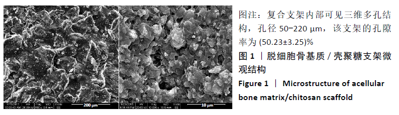
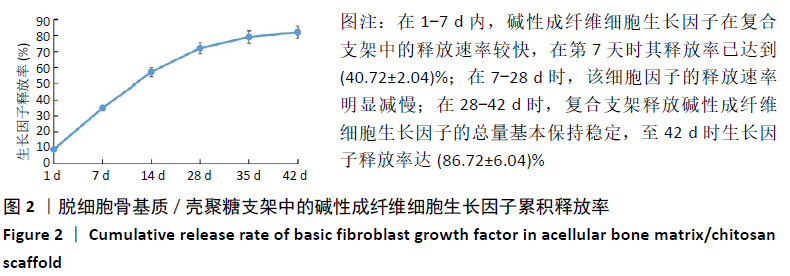
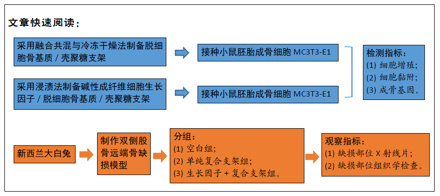
方法:采用融合共混与冷冻干燥法制备脱细胞骨基质/壳聚糖支架,采用浸渍法制备碱性成纤维细胞生长因子/脱细胞骨基质/壳聚糖支架。将小鼠胚胎成骨细胞MC3T3-E1分别接种于两种支架表面,以单独培养的细胞为对照,进行细胞增殖、细胞黏附与成骨基因检测。在36只6月龄新西兰大白兔双侧股骨远端制备直径5 mm的骨缺损模型,抽签法随机分3组,空白组不进行任何干预,单纯复合支架组、生长因子+复合支架组分别植入脱细胞骨基质/壳聚糖支架与碱性成纤维细胞生长因子/脱细胞骨基质/壳聚糖支架,进行骨缺损部位X射线片与组织学检查。
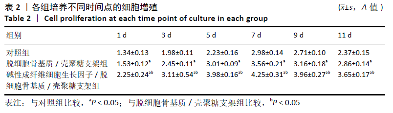
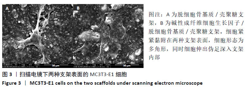
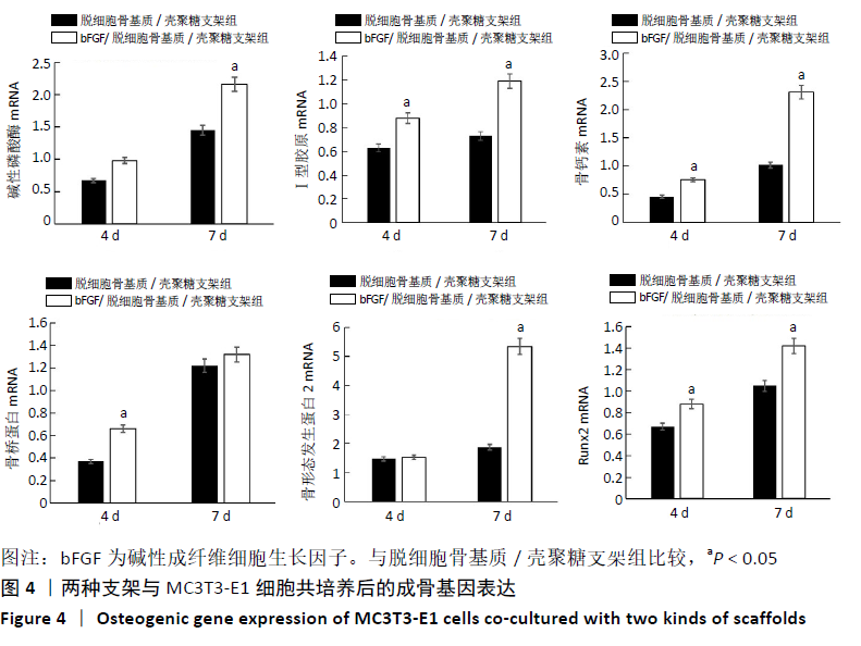
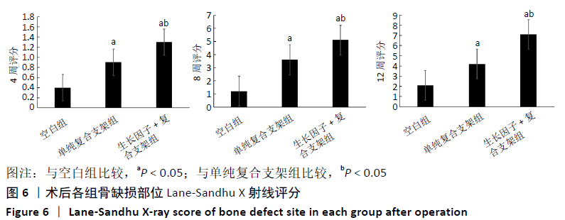
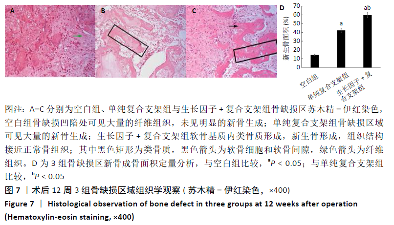
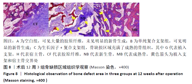
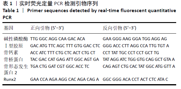
结果与结论:①在培养的1-11 d内,碱性成纤维细胞生长因子/脱细胞骨基质/壳聚糖支架组的细胞增殖快于脱细胞骨基质/壳聚糖支架组   (P < 0.05),脱细胞骨基质/壳聚糖支架组快于对照组(P < 0.05);②细胞与支架共培养4 d后,碱性成纤维细胞生长因子/脱细胞骨基质/壳聚糖支架组培养4,7 d的成骨基因Ⅰ型胶原、骨钙素、骨桥蛋白及Runx2 mRNA表达均高于脱细胞骨基质/壳聚糖支架组(P < 0.05),培养7 d后的碱性磷酸酶mRNA表达高于脱细胞骨基质/壳聚糖支架组(P < 0.05);③共培养7 d后的扫描电镜显示,两组支架均支持MC3T3-E1细胞的黏附;④动物实验X射线片显示,空白组术后12周时无明显的骨修复;单纯复合支架组术后8周时可见新骨生成,术后12周时可见明显新骨生成;细胞因子+复合支架组术后4周时即可见新骨生成,至术后12周时骨缺损部位几乎完全修复;⑤动物实验术后12周的缺损部位苏木精染色与Masson染色显示,空白组可见大量的纤维组织;两支架组可见大量的新骨生成,其中生长因子+复合支架组的新骨生成面积与成熟度高于单纯复合支架组;⑥结果表明,负载碱性成纤维细胞生长因子的脱细胞骨基质/壳聚糖复合支架是较为理想的骨组织修复材料,可促进骨形成。
https://orcid.org/0000-0003-3474-326X (张志文) 
中国组织工程研究杂志出版内容重点:生物材料;骨生物材料口腔生物材料纳米材料缓释材料材料相容性;组织工程

关键词: 碱性成纤维细胞生长因子, 壳聚糖, 支架, 脱细胞骨基质, 复合支架, 股骨缺损修复

Abstract: BACKGROUND: Some studies have shown that basic fibroblast growth factor can promote the proliferation and osteogenic differentiation of mesenchymal stem cells. 
OBJECTIVE: To compare the ability of acellular bone matrix/chitosan scaffold combined with basic fibroblast growth factor in repairing rabbit femoral defect.
METHODS: The acellular bone matrix/chitosan scaffolds were prepared by fusion blending and freeze-drying method, and the acellular bone matrix/chitosan scaffold loaded with basic fibroblast growth factor was prepared by immersion method. Mouse embryonic osteoblasts MC3T3-E1 were seeded on the surface of the two scaffolds for cell proliferation, cell adhesion and osteogenic gene detection. Cells cultured alone served as control. Bone defect models with a diameter of 5 mm were made in bilateral distal femurs using thirty-six 6-month-old New Zealand white rabbits and randomly divided into three groups by drawing lots. The blank group was not intervened. The simple composite scaffold and growth factor + composite scaffold groups were implanted with acellular bone matrix/chitosan scaffold and acellular bone matrix/chitosan scaffold loaded with basic fibroblast growth factor for X-ray and histological examination.
RESULTS AND CONCLUSION: (1) Within 1-11 days of culture, the cell proliferation of basic fibroblast growth factor/acellular bone matrix/chitosan scaffold group was faster than that of the acellular bone matrix/chitosan scaffold group (P < 0.05), and that of the acellular bone matrix/chitosan scaffold group was faster than that of the control group (P < 0.05). (2) After 4 days of co-culture with the scaffold, the mRNA expressions of type I collagen, osteocalcin, osteopontin and Runx2 were higher in the cells in basic fibroblast growth factor/acellular bone matrix/chitosan scaffold group cultured for 4 and 7 days than those of acellular bone matrix/chitosan scaffold group (P < 0.05), and the mRNA expression of alkaline phosphatase was higher than that of acellular bone matrix/chitosan scaffold group after 7 days of culture (P < 0.05). (3) Scanning electron microscopy showed that the scaffolds of the two groups supported the adhesion of MC3T3-E1 cells after 7 days of co-culture. (4) X-ray film of animal experiment showed that there was no obvious bone repair in the blank group at 12 weeks after operation; new bone formation could be seen at 8 weeks after operation in the simple composite scaffold group, and obvious new bone formation could be seen at 12 weeks after operation; new bone formation could be seen at 4 weeks after operation in the cytokine + composite scaffold group, and the bone defect site was almost completely repaired at 12 weeks after operation (5) Hematoxylin staining and Masson staining showed that a large amount of fibrous tissue could be seen in the blank group at 12 weeks after operation; a large amount of new bone formation could be seen in the two scaffold groups, in which the area and maturity of new bone formation in the growth factor + composite scaffold group were higher than those in the simple composite scaffold group. (6) The results showed that the acellular bone matrix/chitosan composite scaffold loaded with basic fibroblast growth factor is an ideal bone tissue repair material, which can promote bone formation.

Key words: basic fibroblast growth factor, chitosan, scaffold, acellular bone matrix, composite scaffold, femoral defect repair

中图分类号: