[1] BIJLSMA JW, BERENBAUM F, LAFEBER FP. Osteoarthritis: an update with relevance for clinical practice. Lancet. 2011;377(9783):2115-2126.
[2] LITWIC A, EDWARDS MH, DENNISON EM, et al. Epidemiology and burden of osteoarthritis. Br Med Bull. 2013;105:185-199.
[3] LIU Q, WANG S, LIN J, et al. The burden for knee osteoarthritis among Chinese elderly: estimates from a nationally representative study. Osteoarthritis Cartilage. 2018;26(12):1636-1642.
4] 王斌,邢丹,林剑浩,等.关节腔内注射治疗膝骨关节炎的研究进展[J].中华关节外科杂志(电子版),2018,12(6):814-820.
[5] 王弘德,李升,陈伟,等.《骨关节炎诊疗指南(2018年版)》膝关节骨关节炎部分的更新与解读[J].河北医科大学学报,2019,40(9): 993-995,1000.
[6] MAKRIS EA, GOMOLL AH, MALIZOS KN, et al. Repair and tissue engineering techniques for articular cartilage. Nat Rev Rheumatol. 2015;11(1):21-34.
[7] 卓群豪,张伟娜,李舰,等. 膝关节腔内注射Sox9转染骨髓间充质干细胞修复膝关节骨关节炎[J].中国组织工程研究,2017,21(5): 736-741.
[8] AL FAQEH H, NOR HAMDAN BM, CHEN HC, et al. The potential of intra-articular injection of chondrogenic-induced bone marrow stem cells to retard the progression of osteoarthritis in a sheep model. Exp Gerontol. 2012;47(6):458-464.
[9] HALEEM AM, SINGERGY AA, SABRY D, et al. The Clinical Use of Human Culture-Expanded Autologous Bone Marrow Mesenchymal Stem Cells Transplanted on Platelet-Rich Fibrin Glue in the Treatment of Articular Cartilage Defects: A Pilot Study and Preliminary Results. Cartilage. 2010;1(4):253-261.
[10] WAKITANI S, OKABE T, HORIBE S, et al. Safety of autologous bone marrow-derived mesenchymal stem cell transplantation for cartilage repair in 41 patients with 45 joints followed for up to 11 years and 5 months. J Tissue Eng Regen Med. 2011;5(2):146-150.
[11] UCCELLI A, MORETTA L, PISTOIA V. Mesenchymal stem cells in health and disease. Nat Rev Immunol. 2008;8(9):726-736.
[12] PITTENGER MF, MACKAY AM, BECK SC, et al. Multilineage potential of adult human mesenchymal stem cells. Science. 1999;284(5411): 143-147.
[13] 李凡,杜联芳.低强度脉冲超声介导骨髓间充质干细胞移植的研究进展[J].中华超声影像学杂志,2017,26(8):729-732.
[14] 于海生,陈文直,王嫣,等.低强度脉冲超声对体外培养的骨髓间充质干细胞增殖效应的研究[J].中国医科大学学报,2011,40(11):
971-974.
[15] 罗显文,李明星.低强度脉冲超声可缓解膝骨关节炎疼痛与修复关节软骨损伤[J].中国组织工程研究,2019,23(3):348-353.
[16] HAYAMI T, PICKARSKI M, ZHUO Y, et al. Characterization of articular cartilage and subchondral bone changes in the rat anterior cruciate ligament transection and meniscectomized models of osteoarthritis. Bone. 2006;38(2):234-243.
[17] MANKIN HJ, JOHNSON ME, LIPPIELLO L. Biochemical and metabolic abnormalities in articular cartilage from osteoarthritic human hips. III. Distribution and metabolism of amino sugar-containing macromolecules. J Bone Joint Surg Am. 1981;63(1):131-139.
[18] LOESER RF, GOLDRING SR, SCANZELLO CR, et al. Osteoarthritis: a disease of the joint as an organ. Arthritis Rheum. 2012;64(6): 1697-1707.
[19] GUPTA PK, DAS AK, CHULLIKANA A, et al. Mesenchymal stem cells for cartilage repair in osteoarthritis. Stem Cell Res Ther. 2012;3(4):25.
[20] KUSUYAMA J, HWAN SEONG C, OHNISHI T, et al. Low-Intensity Pulsed Ultrasound (LIPUS) Stimulation Helps to Maintain the Differentiation Potency of Mesenchymal Stem Cells by Induction in Nanog Protein Transcript Levels and Phosphorylation. J Orthop Trauma. 2016;30(8):
S4-5.
[21] NISHIMORI M, DEIE M, KANAYA A, et al. Repair of chronic osteochondral defects in the rat. A bone marrow-stimulating procedure enhanced by cultured allogenic bone marrow mesenchymal stromal cells. J Bone Joint Surg Br. 2006;88(9):1236-1244.
[22] WAKITANI S, GOTO T, PINEDA SJ, et al. Mesenchymal cell-based repair of large, full-thickness defects of articular cartilage. J Bone Joint Surg Am. 1994;76(4):579-592.
[23] MURPHY JM, FINK DJ, HUNZIKER EB, et al. Stem cell therapy in a caprine model of osteoarthritis. Arthritis Rheum. 2003;48(12): 3464-3474.
[24] VEGA A, MARTÍN-FERRERO MA, DEL CANTO F, et al. Treatment of Knee Osteoarthritis With Allogeneic Bone Marrow Mesenchymal Stem Cells: A Randomized Controlled Trial. Transplantation. 2015;99(8):1681-1690.
[25] CHOI JW, CHOI BH, PARK SH, et al. Mechanical stimulation by ultrasound enhances chondrogenic differentiation of mesenchymal stem cells in a fibrin-hyaluronic acid hydrogel. Artif Organs. 2013;37(7):648-655.
[26] GAO Q, WALMSLEY AD, COOPER PR, et al. Ultrasound Stimulation of Different Dental Stem Cell Populations: Role of Mitogen-activated Protein Kinase Signaling. J Endod. 2016;42(3):425-431.
[27] WANG Y, PENG W, LIU X, et al. Study of bilineage differentiation of human-bone-marrow-derived mesenchymal stem cells in oxidized sodium alginate/N-succinyl chitosan hydrogels and synergistic effects of RGD modification and low-intensity pulsed ultrasound. Acta Biomater. 2014;10(6):2518-2528.
[28] WEI FY, LEUNG KS, LI G, et al. Low intensity pulsed ultrasound enhanced mesenchymal stem cell recruitment through stromal derived factor-1 signaling in fracture healing. PLoS One. 2014;9(9):e106722.
[29] XIA P, WANG X, QU Y, et al. TGF-β1-induced chondrogenesis of bone marrow mesenchymal stem cells is promoted by low-intensity pulsed ultrasound through the integrin-mTOR signaling pathway. Stem Cell Res Ther. 2017;8(1):281.
[30] YANG C, YOU D, HUANG J, et al. Effects of AURKA-mediated degradation of SOD2 on mitochondrial dysfunction and cartilage homeostasis in osteoarthritis. J Cell Physiol. 2019;234(10):17727-17738.
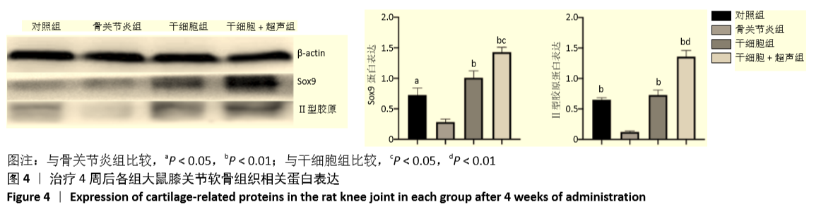
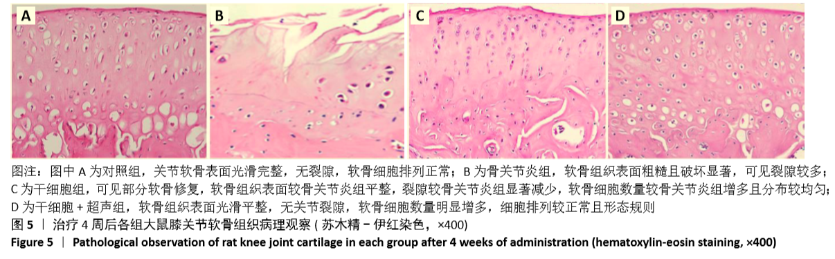
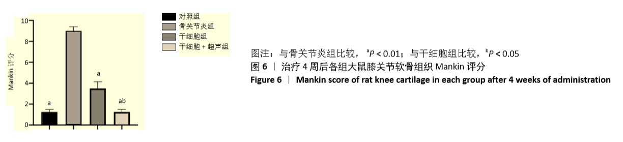
|