中国组织工程研究 ›› 2021, Vol. 25 ›› Issue (28): 4480-4484.doi: 10.12307/2021.061

• 组织工程口腔材料 tissue-engineered oral materials • 上一篇    下一篇

多余树脂粘接剂清除方法对不同边缘间隙全瓷冠边缘完整性的影响

张海洋1,刘建彰2,阚  娜3,李红霞4,王俊丰5,姜海巍1,邱澄宇1,肖  震1   

  1. 1齐齐哈尔市第一医院口腔修复科,黑龙江省齐齐哈尔市  161000;2北京大学口腔医学院口腔修复科,北京市  100081;3连云港市妇幼保健院,江苏省连云港市  222000;4北京瑞泰口腔医院综合二科,北京市 100191;5秦皇岛市第二医院口腔科,河北省秦皇岛市    066000
  • 收稿日期:2020-07-31 修回日期:2020-08-11 接受日期:2020-09-15 出版日期:2021-10-08 发布日期:2021-05-19
  • 通讯作者: Liu Jianzhang, Associate chief physician, Department of Prosthodontics, School of Stomatology, Peking University, Beijing 100081, China
  • 作者简介:张海洋,女,1981年生,黑龙江省齐齐哈尔市人,满族,佳木斯大学口腔医学院硕士,副主任医师,主要从事口腔修复临床研究。
  • 基金资助:
    齐齐哈尔市科学技术局项目(SFZD-2016054),项目负责人:张海洋

Effect of excess resin adhesive removal method on the marginal integrity of all-ceramic crowns with different marginal gaps

Zhang Haiyang1, Liu Jianzhang2, Kan Na3, Li Hongxia4, Wang Junfeng5, Jiang Haiwei1, Qiu Chengyu1, Xiao Zhen1   

  1. 1Department of Prosthodontics, No. 1 Hospital in Qiqihar, Qiqihar 161000, Heilongjiang Province, China; 2Department of Prosthodontics, School of Stomatology, Peking University, Beijing 100081, China; 3Lianyungang Maternal and Child Health Care Hospital, Lianyungang 222000, Jiangsu Province, China; 4Second General Department of Ruitai Stomatology Hospital, Beijing 100191, China; 5Department of Stomatology of Qinhuangdao Second Hospital, Qinhuangdao 066000, Hebei Province, China
  • Received:2020-07-31 Revised:2020-08-11 Accepted:2020-09-15 Online:2021-10-08 Published:2021-05-19
  • Contact: 刘建彰,副主任医师,北京大学口腔医院口腔修复科,北京市 100081
  • About author:Zhang Haiyang, Master, Associate chief physician, Department of Prosthodontics, No. 1 Hospital in Qiqihar, Qiqihar 161000, Heilongjiang Province, China
  • Supported by:
    the Qiqihar Science and Technology Bureau Project, No. SFZD-2016054 (to ZHY)

摘要:

文题释义:
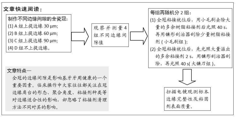
粘接剂清除方法:临床操作过程中在全瓷冠修复体试戴完毕后进行粘接操作时,对于从全瓷冠边缘挤出的多余树脂粘接剂经常使用的两种清除方法是:第一种是先将挤出的大量多余树脂粘接剂用小毛刷刷除后光照40 s,再用镰型刮治器将少量多余粘接剂刮除;另一种是对大量多余树脂粘接剂光照2 s后用镰形刮治器仔细刮除,再光照40 s。实验选取两种不同处理方法旨在找到更适合临床的操作手法,从而指导临床应用。
全瓷冠边缘适应性:是指预备体和修复体边缘的密合程度,是影响修复体临床使用的最重要的因素。不良的边缘密合性会引起粘接剂溶解、发生微渗漏、形成继发龋乃至引发牙髓病及牙周炎症等,从而影响美观及远期修复效果。如果全冠边缘间隙过大、粘接剂缺损等均不利于修复的预后。
背景:国内外有很多关于不同树脂粘接剂、不同肩台形状、不同聚合角度等对全瓷冠边缘完整性影响的研究,但关于多余粘接剂不同处理方法对全瓷冠边缘完整性影响的研究较少。
目的:分析多余树脂粘接剂清除方法对不同边缘间隙全瓷冠边缘完整性的影响。
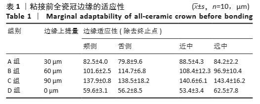
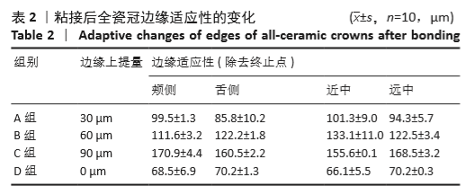
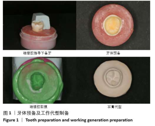
方法:制备40颗离体第三磨牙全瓷冠,随机分成A、B、C、D组,每组10颗。模型扫描完成后,由计算机控制将A、B、C组修复体边缘分别按30,60,90 µm上提,D组不上提边缘,制作Procera氧化铝CAD-CAM全瓷冠。再将A、B、C、D组内随机分2小组,一组全冠粘接就位后,用小毛刷去除大量的多余树脂粘接剂后光照40 s,再用镰形刮治器刮除少量树脂粘接剂(小毛刷组);另一组先光照大量溢出的多余粘接剂2 s,用镰形刮治器刮除,再光照40 s(大镰刀组)。扫描电镜观测标本边缘完整性及粘固剂表面质量。
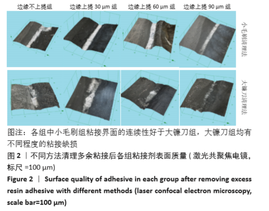
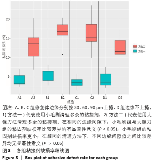
结果与结论:①扫描电镜显示,小毛刷组粘接界面的连续性均较好,镰刀组均有不同程度的粘接缺损;在相边缘间隙下,小毛刷组粘固剂缺损率小于大镰刀组(P < 0.05);在相同粘接剂清除方法下,A、B、C、D组间的粘固剂缺损率比较差异无显著性意义(P﹥0.05);②结果表明,先使用小毛刷去除多余粘接剂后再光照40 s,最后用镰形刮治器去除剩余少量粘接剂的方法更适用于临床。
https://orcid.org/0000-0002-0425-8303 (张海洋) 

中国组织工程研究杂志出版内容重点:生物材料;骨生物材料口腔生物材料纳米材料缓释材料材料相容性;组织工程

关键词: 材料, 口腔, 边缘完整性, 全瓷冠, 粘接剂, 树脂粘接剂, 组织工程, 粘接剂表面质量

Abstract: BACKGROUND: There are many studies on the marginal integrity of all ceramic crowns with different resin adhesives, or different shoulder, different polymerization angles, but there is no research on whether different methods of redundant bonding will affect the marginal integrity of all ceramic crowns. 
OBJECTIVE: To analyze the effect of removing excess resin adhesive on the marginal integrity of all ceramic crowns with different marginal gaps. 
METHODS: Totally 40 extracted third molars were prepared with all ceramic crowns and randomly divided into groups A, B, C, and D (10 teeth in each group). After the model scanning was completed, the edges of restoration in groups A, B and C were lifted at 30, 60 and 90 µm respectively by computer control, while those in group D were not raised. Procera alumina CAD-CAM all ceramic crowns were made. Then, the four groups of groups A, B, C and D were randomly divided into two subgroups. In one subgroup, a large amount of excess resin adhesive was removed with a small brush, and then the light was illuminated for 40 seconds after the full crown bonding. Subsequently, a small amount of resin adhesive was scraped off with sickle scalpel (small brush group). The sample in the other subgroup was exposed to light for 2 seconds, scraped off with sickle scalpel, and then illuminated for 40 seconds (sickle group). The marginal integrity and surface quality of the adhesive were observed by scanning electron microscope.
RESULTS AND CONCLUSION: (1) Scanning electron microscope showed that the continuity of the bonding interface was better in the small brush group. Different degrees of adhesive defects were found in the sickle group. Under the same marginal gap, the defect rate of cement in the small brush group was lower than that in the sickle group (P < 0.05). Under the same adhesive removal method, there was no significant difference in the defect rate of cement between groups A, B, C, and D (P > 0.05). (2) Results indicate that the method of using small brush to remove excess adhesive, then lighting for 40 seconds, and finally using sickle scalpel to remove the remaining small amount of adhesive is more suitable for clinical practice.

Key words: materials, oral cavity, marginal integrity, all ceramic crown, adhesive, resin adhesive, tissue engineering, adhesive surface quality

中图分类号: