中国组织工程研究 ›› 2017, Vol. 21 ›› Issue (36): 5841-5846.doi: 10.3969/j.issn.2095-4344.2017.36.018
• 组织构建实验造模 experimental modeling in tissue construction • 上一篇 下一篇
建立冠状动脉A型病变患者个体化冠状动脉有限元及3D模型
汉 迪,王悦喜,阿 荣,张迎军,王春燕
- 内蒙古医科大学第一附属医院心内科,内蒙古自治区呼和浩特市 010059
Establishment of an individualized three-dimensional finite element model of type A coronary artery lesions
Han Di, Wang Yue-xi, A Rong, Zhang Ying-jun, Wang Chun-yan
- Department of Cardiology, the First Affiliated Hospital of Inner Mongolia Medical University, Hohhot 010059, Inner Mongolia Autonomous Region, China
摘要:
文章快速阅读:
.jpg) 文题释义:
心脏及冠脉有限元模型:利用计算机辅助软件对心脏MRI数据进行处理,模拟建立心脏各功能结构,尤其是冠状动脉,建立后的结构进行测量及分析,了解冠脉有限元模型的特点,模型行3D打印后可以更加直观的进行分析,科学的模型建立为下一步的液固耦合研究及进一步模拟手术提供科学平台。
冠状动脉A型患者液固耦合:利用计算机软件分别对血管及血液进行网格划分,进行耦合计算,分析冠状动脉A型患者狭窄处血流变的特点及耦合各变量的数值。
文题释义:
心脏及冠脉有限元模型:利用计算机辅助软件对心脏MRI数据进行处理,模拟建立心脏各功能结构,尤其是冠状动脉,建立后的结构进行测量及分析,了解冠脉有限元模型的特点,模型行3D打印后可以更加直观的进行分析,科学的模型建立为下一步的液固耦合研究及进一步模拟手术提供科学平台。
冠状动脉A型患者液固耦合:利用计算机软件分别对血管及血液进行网格划分,进行耦合计算,分析冠状动脉A型患者狭窄处血流变的特点及耦合各变量的数值。
文题释义:
心脏及冠脉有限元模型:利用计算机辅助软件对心脏MRI数据进行处理,模拟建立心脏各功能结构,尤其是冠状动脉,建立后的结构进行测量及分析,了解冠脉有限元模型的特点,模型行3D打印后可以更加直观的进行分析,科学的模型建立为下一步的液固耦合研究及进一步模拟手术提供科学平台。
冠状动脉A型患者液固耦合:利用计算机软件分别对血管及血液进行网格划分,进行耦合计算,分析冠状动脉A型患者狭窄处血流变的特点及耦合各变量的数值。摘要
背景:计算机有限元分析可利用患者的MRI影像学资料进行心脏的模拟建模,建立心脏的完整模型,建立好的模型通过计算机处理可行液固耦合,了解冠状动脉的血流变情况。
目的:利用有限元分析软件建立冠状动脉A型病变患者心脏及冠状动脉有限元模型,并进行3D打印,为进一步模拟临床介入手术提供科学依据。
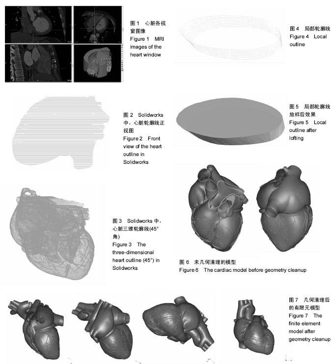
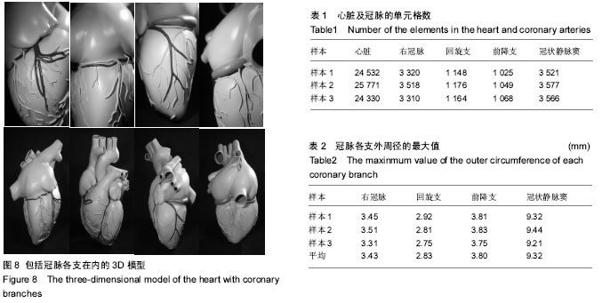
方法:将3例冠状动脉A型病变患者从主动脉弓至心尖行MRI扫描,将图像以Dicom格式导入Mimics 17.0软件中,通过建模并行几何清理,建立完整的包括冠状动脉在内的心脏模型。将三维模型导入Geomagic Studio 11.0软件中,对模型进行进一步图像处理,最后将三维模型导入ANSYS14.0有限元分析软件中,通过附加材料属性建立具有生物性能的有限元模型,并将模型连接3D打印机上行3D打印。
结果与结论:①成功建立了3例冠状动脉A型病变患者包括冠状动脉在内的三维有限元模型,建立的心脏模型,网格划分均为六面体实体单元,单元数分别为24 532,25 771,24 330个,同时建立了冠状动脉各支的模型,右冠脉的单元数分别为3 320,3 518,3 310;回旋支分别为1 148,1 176,1 164;前降支分别为1 025,1 049,1 068,建立完成后的模型成功完成3D打印;②综上,建立了包括冠状动脉在内的心脏三维有限元模型,成功完成3D打印,打印后的冠状动脉形态逼真,可清晰的显示右冠状动脉,前降支、回旋支及冠状静脉窦。模型的建立为临床介入手术的模拟研究搭建了科学的平台,同时冠脉有限元模型的建立为下一步评估血液与血管液固双向耦合分析研究打下了坚实的基础,使其成为血液流变研究一种新的科学方法。
中国组织工程研究杂志出版内容重点:组织构建;骨细胞;软骨细胞;细胞培养;成纤维细胞;血管内皮细胞;骨质疏松;组织工程
ORCID: 0000-0003-1168-0534(汉迪)
中图分类号:
.jpg) 文题释义:
心脏及冠脉有限元模型:利用计算机辅助软件对心脏MRI数据进行处理,模拟建立心脏各功能结构,尤其是冠状动脉,建立后的结构进行测量及分析,了解冠脉有限元模型的特点,模型行3D打印后可以更加直观的进行分析,科学的模型建立为下一步的液固耦合研究及进一步模拟手术提供科学平台。
冠状动脉A型患者液固耦合:利用计算机软件分别对血管及血液进行网格划分,进行耦合计算,分析冠状动脉A型患者狭窄处血流变的特点及耦合各变量的数值。
文题释义:
心脏及冠脉有限元模型:利用计算机辅助软件对心脏MRI数据进行处理,模拟建立心脏各功能结构,尤其是冠状动脉,建立后的结构进行测量及分析,了解冠脉有限元模型的特点,模型行3D打印后可以更加直观的进行分析,科学的模型建立为下一步的液固耦合研究及进一步模拟手术提供科学平台。
冠状动脉A型患者液固耦合:利用计算机软件分别对血管及血液进行网格划分,进行耦合计算,分析冠状动脉A型患者狭窄处血流变的特点及耦合各变量的数值。