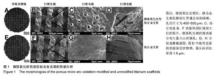
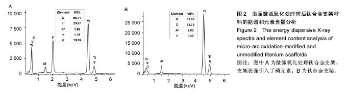
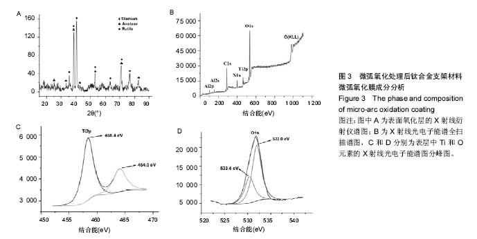
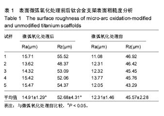
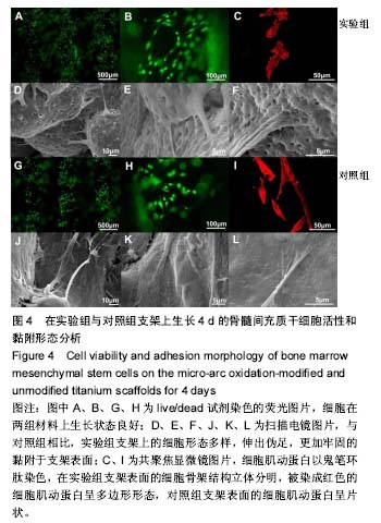
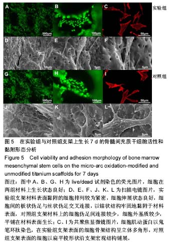
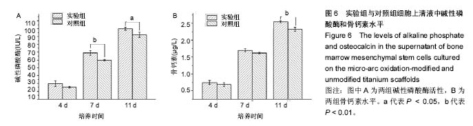
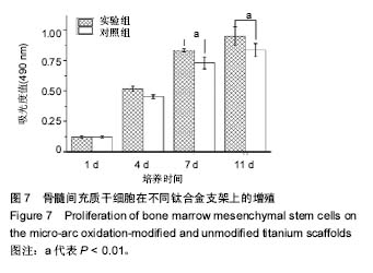
| [1]Uchida M,Oyane A,Kim HM,et al.Biomimetic coating of laminin–apatite composite on titanium metal and its excellent cell-adhesive properties. Adv Mater.2004;37(13):1071-1074.[2]Smith CJ,Derguti F,Hernandez Nava EH, et al.Dimensional accuracy of electron beam melting (EBM) additive manufacture with regard to weight optimized truss structures.J Mater Process Tech.2016;229: 128-138.[3]Jia Z,Li M,Xiu P,et al.A novel cytocompatible, hierarchical porous Ti6Al4V scaffold with immobilized silver nanoparticles.Mater Lett. 2015;157:143-146.[4]Parthasarathy J,Starly B,Raman S,et al.Mechanical evaluation of porous titanium (Ti6Al4V) structures with electron beam melting(EBM).J Mech Behav Biomed Mater. 2010;3:249-259.[5]Li SJ,Murr LE,Cheng XY,et al.Compression fatigue behavior of Ti-6Al-4V mesh arrays fabricated by electron beam melting. Acta Mater.2012;60(3):793-802. [6]刘邦定,郭征,郝玉琳,等.多孔钛合金不同孔径大小对新骨长入的影响[J].现代生物医学进展,2012,12(9):1601-1604. [7]Cremasco A,Messias AD,Esposito AR,et al.Effects of alloying elements on the cytotoxic response of titanium alloys.Mater Sci Eng C. 2011;31(5):833-839. [8]Su Y,Li K,Zhang L,et al.Ca-P bioactive coating prepared by combining microwave-hydrothermal and supersonic atmospheric plasma spraying methods.Mater Sci Eng C. 2017;72:371-377.[9]?erban VA,Ro?u RA,Bucur AI,et al.Deposition of titanium nitride layers by electric arc–reactive plasma spraying method. Appl Surf Sci. 2013; 265:245-249.[10]Laha T,Agarwal A,Mckechnie T,et al.Synthesis and characterization of plasma spray formed carbon nanotube reinforced aluminum composite. Mat Sci Eng A. 2004;381(1): 249-258.[11]Zhang W,Liu W,Liu Y,et al.Tribological behaviors of single and dual sol–gel ceramic films on Ti6Al4V.Ceram Int. 2009;35(4): 1513-1520.[12]Wang XX,Hayakawa S,Tsuru K,et al.Bioactive titania-gel layers formed by chemical treatment of Ti substrate with a H2O2/HCl solution. Biomaterials.2002;23(5):1353-1357.[13]Catauro M,Bollino F,Giovanardi R,et al.Modification of Ti6Al4V implant surfaces by biocompatible TiO2/PCL hybrid layers prepared via sol-gel dip coating: structural characterization, mechanical and corrosion behavior.Mater Sci Eng C.2016;74:501-507.[14]Yan Y,Sun J,Han Y,et al.Microstructure and bioactivity of Ca, P and Sr doped TiO2 coating formed on porous titanium by micro-arc oxidation. Surf Coat Tech.2010;205:1702-1713.[15]Wu J,Liu R,Wang B,et al.Preparation and characterization of carburized layer on pure aluminum by plasma electrolysis. Surf Coat Tech.2015;269:119-124. [16]Durdu S,Ömer Faruk Deniz,Kutbay I,et al.Characterization and formation of hydroxyapatite on Ti6Al4V coated by plasma electrolytic oxidation.J Alloys Compounds.2013;551(5):422-429.[17]Wang Y,Yu H, Chen C,et al.Review of the biocompatibility of micro-arc oxidation coated titanium alloys.Mater Design. 2015;85:640-652.[18]刘帅.钛合金材料表面阳极氧化和微弧氧化改性涂层的构建及生物学研究[D].第四军医大学,2014.[19]Lim YW,Kwon SY,Sun DH,et al.Enhanced cell integration to titanium alloy by surface treatment with micro-arc oxidation: A pilot study.Clin Orthop Relat Res.2009;467(9):2251-2258. [20]Zhu X,Chen J,Scheideler L,et al.Cellular reactions of osteoblasts to micron- and submicron-scale porous structures of titanium surfaces. Cells Tissues Organs.2004;178(1):13-22.[21]Yu SC,Park SN,Suh H.Adipose tissue engineering using mesenchymal stem cells attached to injectable PLGA spheres. Biomaterials. 2005; 26(29):5855-5863.[22]卢宁,赵龙凤,李红,等.应用全骨髓贴壁法获取高纯度大鼠骨髓间充质干细胞的实验研究[J].山西医科大学学报,2010,41(3): 277-280.[23]Qiao LP,Lou J,Zhang SF,et al.The entrance mechanism of calcium and phosphorus elements into micro-arc oxidation coatings developed on Ti6Al4V alloy.Surf Coat Tech.2015; 285:187-196.[24]Zhang RF,Qiao LP,Qu B,et al.Biocompatibility of micro-arc oxidation coatings developed on Ti6Al4V alloy in a solution containing organic phosphate.Mater Lett.2015;153(9):77-80.[25]方丽茹,翁文剑,沈鸽,等.骨组织工程支架及生物材料研究[J].生物医学工程学杂志, 2003,20(1):148-152.[26]Tsai MT,Chang YY,Huang HL,et al.Micro-arc oxidation treatment enhanced the biological performance of human osteosarcoma cell line and human skin fibroblasts cultured on titanium–zirconium films. Surf Coat Tech.2016;303:268-276.[27]李玉梅,查年保,马威,等. 微弧氧化处理对钛表面MC3T3-E1细胞生物学行为的影响[J].上海口腔医学, 2015,24(5):551-556.[28]Huang Y,Wang Y,Ning C,et al.Hydroxyapatite coatings produced on commercially pure titanium by micro-arc oxidation.Biomed Mater. 2007;2(3):196-201.[29]Cheng XY,Li SJ,Murr LE,et al.Compression deformation behavior of Ti-6Al-4V alloy with cellular structures fabricated by electron beam melting.J Mech Behav Biomed Mater. 2012;16(2):153-162.[30]Li XK,Yuan CF,Wang JL,et al.The treatment effect of porous titanium alloy rod on the early stage talar osteonecrosis of sheep.PLoS One. 2013;8(3):e58459.[31]吴穗丹,王焱,张辉,等.钛表面微弧氧化涂层的细胞生物活性[J].中国组织工程研究, 2013,17(47):8169-8174.[32]Zhao Z,Chen X,Chen A,et al.Synthesis of bioactive ceramic on the titanium substrate by micro-arc oxidation.J Biomed Mater Res A. 2009;90(2):438-445.[33]Teng FY,Ko CL,Kuo HN,et al.A comparison of epithelial cells, fibroblasts, and osteoblasts in dental implant titanium topographies. Bioinorg Chem Appl.2012;2012(4):687291.[34]Krupa D,Baszkiewicz J,Zdunek J,et al. Effect of plasma electrolytic oxidation in the solutions containing Ca, P, Si, Na on the properties of titanium.J Biomed Mater Res B Appl Biomater.2012;100: 2156-2166.[35]Zhou R,Wei D,Feng W,et al.Bioactive coating with hierarchical double porous structure on titanium surface formed by two-step micro-arc oxidation treatment.Surf Coat Tech.2014;252(9):148-156.[36]Erfanifar E,Aliofkhazraei M,Nabavi HF,et al.Growth kinetics and morphology of microarc oxidation coating on titanium. Surf Coat Tech. 2017;315:567-576. [37]St-Pierre JP,Gauthier M,Lefebvre LP,et al.Three-dimensional growth of differentiating MC3T3-E1 pre-osteoblasts on porous titanium scaffolds. Biomaterials.2005;26(35):7319-7328.[38]Benoit DS,Schwartz MP,Durney AR,et al.Small functional groups for controlled differentiation of hydrogel-encapsulated human mesenchymal stem cells.Nat Mater.2008;7(10): 816-823.[39]郭宝刚,梁军,陈建敏,等.氧化时间对Ti6Al4V微弧氧化膜结构与性能的影响[J].中国有色金属学报,2005,15(6):981-986.[40]Curran JA,Clyne TW.Porosity in plasma electrolytic oxide coatings. Acta Mater.2006; 54(7):1985-1993.[41]Chen HT,Hsiao CH,Long HY,et al.Micro-arc oxidation of β-titanium alloy: structural characterization and osteoblast compatibility.Surf Coat Tech.2009;204(6):1126-1131.[42]Kunzler TP,Huwiler C,Drobek T,et al.Systematic study of osteoblast response to nanotopography by means of nanoparticle-density gradients.Biomaterials.2007;28(33): 5000-5006.[43]Mizokami A,Kawakubo-yasukochi T,Hirata M.Osteocalcin and its endocrine functions.Biochem Pharmacol.2017;132:1-8.[44]赵煜,李冰雁,杜莉,等.磁性附着体模拟静磁场对成骨细胞分化功能的影响[J].中华口腔医学研究,2011,05(2):151-155.[45]丁思阳,夏露,陈宁,等.微弧氧化纯钛表面对成骨细胞蛋白合成功能的影响[J].口腔医学研究,2012,28(2):125-128. |
.jpg)
.jpg)