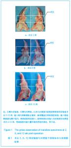
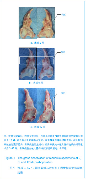
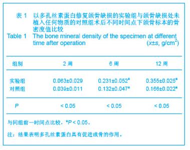
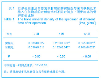
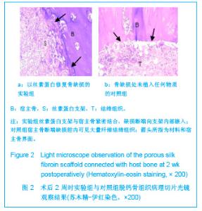
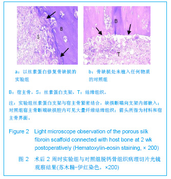
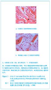
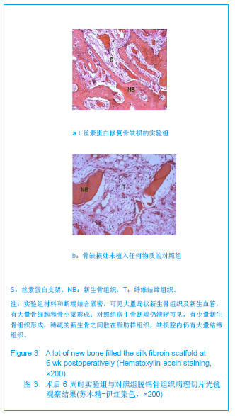
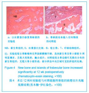
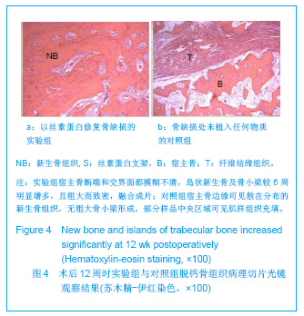
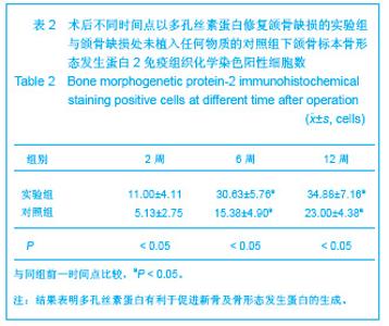
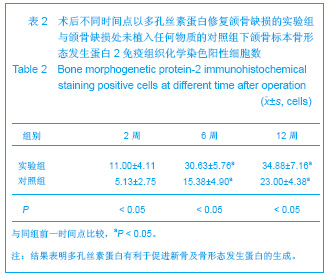
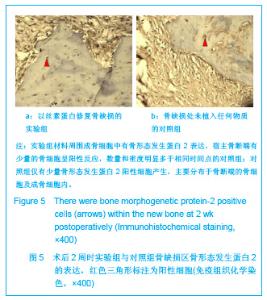
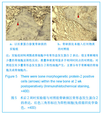
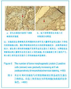
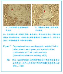
| [1] Gazdag AR,Lane JM,Glaser D,et al.Alternatives to Autogenous Bone Graft: Efficacy and Indications.J Am Acad Orthop Surg.1995;3(1): 1-8.[2] Meinel L,Betz O,Fajardo R,et al.Silk based biomaterials to heal critical sized femur defects.Bone.2006;39(4): 922-931.[3] Ebraheim NA,Elgafy H,Xu R. Bone-graft harvesting from iliac and fibular donor sites: techniques and complications.J Am Acad Orthop Surg.2001; 9(3): 210-218.[4] Ning L,Xue M,Huang HN,et al.Zhongguo Xiufu Chongjian Waike Zazhi. 2000; 14(1): 44-48. 宁丽,薛淼,黄海宁,等.皮肤再生膜的生物相容性系列研究[J].中国修复与重建外科杂志,2000,14(1): 44-48.[5] Meinel L,Hofmann S,Karageorgiou V,et al.The inflammatory responses to silk films in vitro and in vivo.Biomaterials. 2005; 26(2):147-155.[6] Wang Y,Rudym DD,Walsh A,et al.In vivo degradation of three-dimensional silk fibroin scaffolds.Biomaterials.2008; 29(24-25):3415-3428.[7] Sofia S,McCarthy MB,Gronowicz G,et al.Functionalized silk-based biomaterials for bone formation.J Biomed Mater Res.2001; 54(1): 139-148.[8] Wang Y,Blasioli DJ,Kim HJ,et al.Cartilage tissue engineering with silk scaffolds and human articular chondrocytes. Biomaterials.2006;27(25): 4434-4442.[9] Tamada Y.New process to form a silk fibroin porous 3-D structure. Biomacromolecules.2005; 6(6): 3100-3106.[10] Mauney JR,Nguyen T,Gillen K,et al.Engineering adipose-like tissue in vitro and in vivo utilizing human bone marrow and adipose-derived mesenchymal stem cells with silk fibroin 3D scaffolds.Biomaterials.2007; 28(35): 5280-5290.[11] The Ministry of Science and Technology of the People’s Republic of China. Guidance suggestion of caring laboratory animals.2006-09-30. 中华人民共和国科学技术部.关于善待实验动物的指导性意见. 2006-09-30.[12] Schmitz JP,Hollinger JO.The critical size defect as an experimental model for craniomandibulofacial nonunions.Clin Orthop Relat Res.1986;(205): 299-308.[13] Hollinger JO,Kleinschmidt JC.The critical size defect as an experimental model to test bone repair materials.J Craniofac Surg.1990; 1(1): 60-68.[14] Langer R,Vacanti JP. Tissue engineering.Science.1993; 260(5110): 920-926.[15] Shimizu Y. Tissue engineering for soft tissue.In: The Tissue Engineering for Therapeutic Use, Ikada Y, Enomoto S, Eds. Elsevier Science: Amsterdam. 1998: 119-120.[16] Salgado AJ,Coutinho OP,Reis RL. Bone tissue engineering: state of the art and future trends. Macromol Biosci.2004; 4(8): 743-765.[17] Burg KJ,Porter S,Kellam JF.Biomaterial developments for bone tissue engineering. Biomaterials.2000; 21(23): 2347-2359.[18] Zhang YG,Lu SB,Wang JF.Zhonghua Waike Zazhi. 1998;18(11): 678-681. 张永刚,卢世璧,王继芳.利用隔膜技术探讨骨折不愈合原因[J].中华外科杂志,1998,18(11): 678-681. |