Chinese Journal of Tissue Engineering Research ›› 2021, Vol. 25 ›› Issue (20): 3144-3149.doi: 10.3969/j.issn.2095-4344.3188
Previous Articles Next Articles
Correlation between skeletal muscle fiber characteristics and bone mineral density in postmenopausal women with hip fractures
Lin Haishan, Mieralimu Muertizha, Li Peng, Ma Chao, Wang Li
- Geriatric Ward of Orthopedics, People’s Hospital of Xinjiang Uygur Autonomous Region, Urumqi 830001, Xinjiang Uygur Autonomous Region, China
-
Received:2020-04-07Revised:2020-04-11Accepted:2020-06-12Online:2021-07-18Published:2021-01-15 -
Contact:Wang Li, Chief physician, Geriatric Ward of Orthopedics, People’s Hospital of Xinjiang Uygur Autonomous Region, Urumqi 830001, Xinjiang Uygur Autonomous Region, China -
About author:Lin Haishan, Master candidate, Physician, Geriatric Ward of Orthopedics, People’s Hospital of Xinjiang Uygur Autonomous Region, Urumqi 830001, Xinjiang Uygur Autonomous Region, China -
Supported by:the Natural Science Foundation of Xinjiang Uygur Autonomous Region (General Project), No. 2017D01C127 (to WL)
CLC Number:
Cite this article
Lin Haishan, Mieralimu Muertizha, Li Peng, Ma Chao, Wang Li. Correlation between skeletal muscle fiber characteristics and bone mineral density in postmenopausal women with hip fractures[J]. Chinese Journal of Tissue Engineering Research, 2021, 25(20): 3144-3149.
share this article
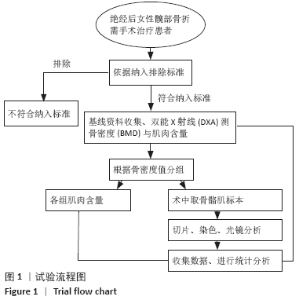
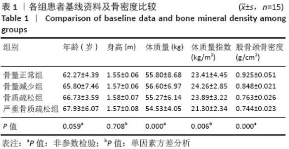
2.1 参与者数量分析 纳入患者60例,分为4组,治疗过程无脱失,全部进入结果分析。 2.2 患者基线资料比较 各组研究对象体质量、体质量指数、股骨颈骨密度比较差异有显著性意义(P < 0.05),但各组间年龄、身高比较差异无显著性意义(P > 0.05),见表1。试验流程图见图1。"

"

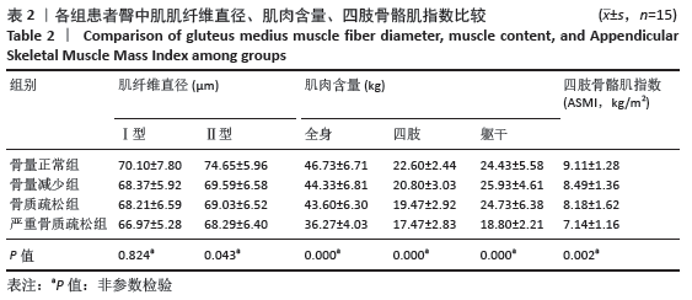
2.3 臀中肌肌纤维直径、肌肉含量及四肢骨骼肌指数比较 各组Ⅰ型肌纤维直径大小分布差异无显著性意义(P > 0.05),各组Ⅱ型肌纤维直径大小、全身、四肢、躯干肌肉含量及ASMI分布组间差异有显著性意义(P < 0.05),见表2。"
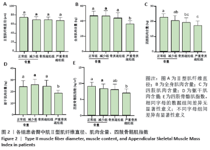
组间多重比较提示:Ⅱ型肌纤维直径差异无显著性意义(P > 0.05),见图2A;全身肌肉含量与躯干肌肉含量:严重骨质疏松组显著低于其余3组(P < 0.05),但其余3组两两比较差异无显著性意义(P > 0.05);四肢肌肉含量:骨量正常组显著高于骨质疏松组、严重骨质疏松组(P < 0.05),骨量减少组与严重骨质疏松组比较差异有显著性意义(P < 0.05);四肢骨骼肌指数:严重骨质疏松组与骨量正常组、骨量减少组比较差异有显著性意义(P < 0.05),见图2B-E。"

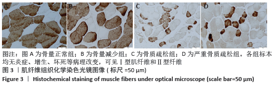
2.4 肌纤维组织化学染色观察 常规组织化学染色显示,所有标本镜下均无炎症、增生、坏死等病理改变,可见Ⅰ型肌纤维和Ⅱ型纤维,见图3。"
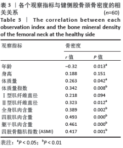
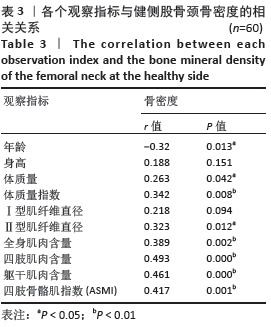
2.5 骨密度与各个观察指标的相关关系 股骨颈骨密度与年龄、体质量、Ⅱ型肌纤维直径相关系数对应的P值均< 0.05,说明其与年龄、体质量、Ⅱ型肌纤维直径具有相关性,其中年龄呈负相关,体质量及Ⅱ型肌纤维直径呈正相关;股骨颈骨密度与体质量指数、全身肌肉含量、四肢肌肉含量、躯干肌肉含量、ASMI相关系数对应的P值< 0.05,说明上述指标与骨密度具有显著相关性;与身高、Ⅰ型肌纤维直径相关系数对应P值均> 0.05,说明上述指标与骨密度不具有相关关系,见表3。"
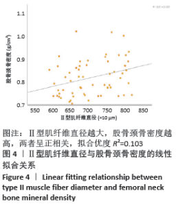
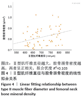
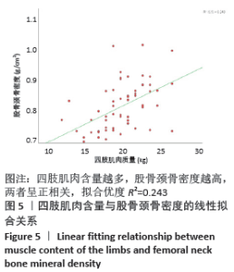
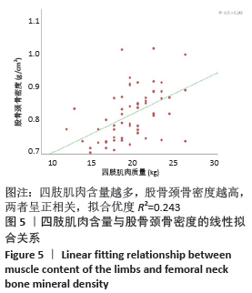
2.6 Ⅱ型肌纤维直径、四肢肌肉含量与股骨颈骨密度的关系 表3中Ⅱ型肌纤维直径及四肢肌肉含量所对应相关系数r分别为:0.323,0.493,与股骨颈骨密度相关性较强,故进一步分析二者分别与股骨颈骨密度的线性关系。Ⅱ型肌纤维直径越大,股骨颈骨密度越高,两者呈正相关,拟合优度R2=0.103,见图4;四肢肌肉含量越多,股骨颈骨密度越高,两者呈正相关,拟合优度R2=0.243,见图5。"
"
| [1] DIAB DL, WATTS NB. Postmenopausal osteoporosis.Curr Opin Endocrinol Diabetes Obes. 2013;20(6):501-509. [2] LESLIE WD, MORIN SN. Osteoporosis epidemiology 2013: implications for diagnosis, risk assessment, and treatment. Curr Opin Rheumatol. 2014;6:440-446. [3] MORIWAKI S, SUZUKI K, MURAMATSU M, et al. Delphinidin, one of the major anthocyanidins, prevents bone loss through the inhibition of excessive osteoblastogenesis in osteoporosis model mice. PLoS One. 2014;9:e97177. [4] EASTELL R, BLACK DM, BOONEN S, et al. HORIZON Pivotal Fracture Trial. Effect of once-yearly zoledronic acid five milligrams on fracture risk and change in femoral neck bone mineral density. J Clin Endocrinol Metab. 2009;94:3215-3225. [5] PISCITELLI P, BRANDI M, CAWSTON H, et al. Epidemiological burden of postmenopausal osteoporosis in Italy from 2010 to 2020: estimations from a disease model. Calcif Tissue Int. 2014;95:419-427. [6] 杨丽君,吴永华,张俐.肌少症、骨质疏松症的关系及研究进展[J].中国骨质疏松杂志,2017,23(8):1112-1116. [7] MONACO MD, VALLERO F, MONACO RD, et al. Prevalence of sarcopenia and its association with osteoporosis in 313 older women following a hip fracture. Arch Gerontol Geriatr. 2011;52(1):71-74. [8] HWANG JA, KIM YS, LEEM AY, et al. Clinical implications of sarcopenia on decreased bone density in men with COPD.Chest. 2017;151(5): 1018-1027. [9] BINKLEY N, BUEHRING B. Beyond FRAX:It’s time to consider “Sarco-Osteopenia”. J Clin Densitom. 2009;12(4):413-416. [10] TERRACCIANO C, CELI M, LECCE D, et al. Differential features of muscle fiber atrophy in osteoporosis and osteoarthritis. Osteoporos Int. 2013; 24(3):1095‐1100. [11] 章涛,张潜,胡锡阶,等.改良骨骼肌肌纤维分型ATP酶染色方法[J].西安交通大学学报(医学版),2009,30(2):254-256. [12] BROOKE MH, KAISER KK. Muscle fiber types: how many and what kind?. Arch Neurol. 1970;23(4):369-379. [13] 中华医学会骨质疏松和骨矿盐疾病分会.肌少症共识[J].中华骨质疏松和骨矿盐疾病杂志,2016,9(3):215-227. [14] MAUREL DB, JAHN K, LARA-CASTILLO N. Muscle-bone crosstalk: Emerging opportunities for novel therapeutic approaches to treat musculoskeletal pathologies. Biomedicines. 2017;5(4):62. [15] YOO JI, HA YC, KWON HB, et al. High Prevalence of Sarcopenia in Korean Patients after Hip Fracture: a Case-Control Study.J Korean Med Sci. 2016;31(9):1479-1484. [16] 黄宏兴,吴青,李跃华,等.肌肉、骨骼与骨质疏松专家共识[J].中国骨质疏松杂志,2016,22(10):1221-1229,1236. [17] 何跃辉,陈狄,高谦,等.围绝经期和绝经后女性骨密度的变化及其相关危险因素分析[J].中国骨质疏松杂志,2019,25(2):185-188, 211. [18] 杜艳萍,朱汉民. 肌少症的诊疗和防治研究[J].中华骨质疏松和骨矿盐疾病杂志,2014,7(1):1-8. [19] JOANISSE S, NEDERVEEN JP, SNIJDERS T, et al. Skeletal Muscle Regeneration, Repair and Remodelling in Aging: The Importance of Muscle Stem Cells and Vascularization. Gerontology. 2017;63(1):91‐100. [20] SAKUSHIMA K, YOSHIKAWA M, OSAKI T, et al. Moderate hypoxia promotes skeletal muscle cell growth and hypertrophy in C2C12 cells.Biochem Biophys Res Commun. 2020;525(4):921-927. [21] CHAILLOU T, SANNA I, KADI F. Glutamine-stimulated in vitro hypertrophy is preserved in muscle cells from older women. Mech Ageing Dev. 2020;187:111228. [22] SARCHIELLI E, COMEGLIO P, FILIPPI S, et al. Testosterone improves muscle fiber asset and exercise performance in a metabolic syndrome model. J Endocrinol. 2020;245(2):259-279. [23] WRIGHT NC, LOOKER AC, SAAG KG, et al. The recent prevalence of osteoporosis and low bone mass in the United States based on bone mineral density at the femoral neck or lumbar spine. J Bone Miner Res. 2014;29(11):2520-2526. [24] SANG JK, YANG WG, CHO E, et al. Relationship between weight, body mass index and bone mineral density of lumbar spine in women. J Bone Metab. 2012;19(2):95-102. [25] CHAN MY, FROST SA, CENTER JR, et al. Relationship between body mass index and fracture risk is mediated by bone mineral density. J Bone Miner Res. 2014;29(11):2327-2335. [26] KOENDERS MI, VAN DEN BERG WB. Novel therapeutic targets inrheumatoid arthritis. Trends Pharmacol Sci. 2015;36:89-195. [27] PAPANICOLAOU DA, ATHER SN, ZHU H, et al. A phase IIA randomized, placebo-controlled clinical trial to study the efficacy and safety of the selective androgen receptor modulator (SARM), MK-0773 in female participants with sarcopenia. J Nutr Health Aging. 2013;17(6):533‐543. [28] LAMONTE MJ, WACTAWSKI-WENDE J, LARSON JC, et al. Association of Physical Activity and Fracture Risk Among Postmenopausal Women.JAMA Netw Open. 2019;2(10):e1914084. [29] HARBER MP, KONOPKA AR, UNDEM MK, et al. Aerobic exercise training induces skeletal muscle hypertrophy and age-dependent adaptations in myofiber function in young and older men. J Appl Physiol (1985). 2012;113(9):1495‐1504. [30] Erlich AT, Tryon LD, Crilly MJ, et al. Function of specialized regulatory proteins and signaling pathways in exercise-induced muscle mitochondrial biogenesis. Integr Med Res. 2016;5(3):187‐197. [31] 刘建,刘杰,丁九阳,等.低氧高糖条件下家兔骨骼肌Ⅰ型纤维向Ⅱ型纤维转化的作用[J].局解手术学杂志,2019,28(2):99-103. [32] Wakabayashi H, Sakuma K. Rehabilitation nutrition for sarcopenia with disability: a combination of both rehabilitation and nutrition care management. J Cachexia Sarcopenia Muscle. 2014;5(4):269‐277. [33] Martone AM, Lattanzio F, Abbatecola AM, et al. Treating sarcopenia in older and oldest old. Curr Pharm Des. 2015;21(13): 1715‐1722. |
| [1] | Hu Kai, Qiao Xiaohong, Zhang Yonghong, Wang Dong, Qin Sihe. Treatment of displaced intra-articular calcaneal fractures with cannulated screws and plates: a meta-analysis of 15 randomized controlled trials [J]. Chinese Journal of Tissue Engineering Research, 2021, 25(9): 1465-1470. |
| [2] | Huang Dengcheng, Wang Zhike, Cao Xuewei. Comparison of the short-term efficacy of extracorporeal shock wave therapy for middle-aged and elderly knee osteoarthritis: a meta-analysis [J]. Chinese Journal of Tissue Engineering Research, 2021, 25(9): 1471-1476. |
| [3] | Xu Feng, Kang Hui, Wei Tanjun, Xi Jintao. Biomechanical analysis of different fixation methods of pedicle screws for thoracolumbar fracture [J]. Chinese Journal of Tissue Engineering Research, 2021, 25(9): 1313-1317. |
| [4] | Jiang Yong, Luo Yi, Ding Yongli, Zhou Yong, Min Li, Tang Fan, Zhang Wenli, Duan Hong, Tu Chongqi. Von Mises stress on the influence of pelvic stability by precise sacral resection and clinical validation [J]. Chinese Journal of Tissue Engineering Research, 2021, 25(9): 1318-1323. |
| [5] | Zhang Tongtong, Wang Zhonghua, Wen Jie, Song Yuxin, Liu Lin. Application of three-dimensional printing model in surgical resection and reconstruction of cervical tumor [J]. Chinese Journal of Tissue Engineering Research, 2021, 25(9): 1335-1339. |
| [6] | Zhang Yu, Tian Shaoqi, Zeng Guobo, Hu Chuan. Risk factors for myocardial infarction following primary total joint arthroplasty [J]. Chinese Journal of Tissue Engineering Research, 2021, 25(9): 1340-1345. |
| [7] | Wei Wei, Li Jian, Huang Linhai, Lan Mindong, Lu Xianwei, Huang Shaodong. Factors affecting fall fear in the first movement of elderly patients after total knee or hip arthroplasty [J]. Chinese Journal of Tissue Engineering Research, 2021, 25(9): 1351-1355. |
| [8] | Wang Jinjun, Deng Zengfa, Liu Kang, He Zhiyong, Yu Xinping, Liang Jianji, Li Chen, Guo Zhouyang. Hemostatic effect and safety of intravenous drip of tranexamic acid combined with topical application of cocktail containing tranexamic acid in total knee arthroplasty [J]. Chinese Journal of Tissue Engineering Research, 2021, 25(9): 1356-1361. |
| [9] | Xiao Guoqing, Liu Xuanze, Yan Yuhao, Zhong Xihong. Influencing factors of knee flexion limitation after total knee arthroplasty with posterior stabilized prostheses [J]. Chinese Journal of Tissue Engineering Research, 2021, 25(9): 1362-1367. |
| [10] | Huang Zexiao, Yang Mei, Lin Shiwei, He Heyu. Correlation between the level of serum n-3 polyunsaturated fatty acids and quadriceps weakness in the early stage after total knee arthroplasty [J]. Chinese Journal of Tissue Engineering Research, 2021, 25(9): 1375-1380. |
| [11] | Zhang Chong, Liu Zhiang, Yao Shuaihui, Gao Junsheng, Jiang Yan, Zhang Lu. Safety and effectiveness of topical application of tranexamic acid to reduce drainage of elderly femoral neck fractures after total hip arthroplasty [J]. Chinese Journal of Tissue Engineering Research, 2021, 25(9): 1381-1386. |
| [12] | Wang Haiying, Lü Bing, Li Hui, Wang Shunyi. Posterior lumbar interbody fusion for degenerative lumbar spondylolisthesis: prediction of functional prognosis of patients based on spinopelvic parameters [J]. Chinese Journal of Tissue Engineering Research, 2021, 25(9): 1393-1397. |
| [13] | Lü Zhen, Bai Jinzhu. A prospective study on the application of staged lumbar motion chain rehabilitation based on McKenzie’s technique after lumbar percutaneous transforaminal endoscopic discectomy [J]. Chinese Journal of Tissue Engineering Research, 2021, 25(9): 1398-1403. |
| [14] | Chen Xinmin, Li Wenbiao, Xiong Kaikai, Xiong Xiaoyan, Zheng Liqin, Li Musheng, Zheng Yongze, Lin Ziling. Type A3.3 femoral intertrochanteric fracture with augmented proximal femoral nail anti-rotation in the elderly: finite element analysis of the optimal amount of bone cement [J]. Chinese Journal of Tissue Engineering Research, 2021, 25(9): 1404-1409. |
| [15] | Du Xiupeng, Yang Zhaohui. Effect of degree of initial deformity of impacted femoral neck fractures under 65 years of age on femoral neck shortening [J]. Chinese Journal of Tissue Engineering Research, 2021, 25(9): 1410-1416. |
| Viewed | ||||||
|
Full text |
|
|||||
|
Abstract |
|
|||||