| [1]McCulloch C A,Bordin S. Role of fibroblast subpopulations in periodontal physiology and pathology. J Periodontal Res. 1991; 26(3 Pt 1): 144-154.
[2]Bartold PM, Shi S, Gronthos S. Stem cells and periodontal regeneration. Periodontol. 2000;40: 164-172.
[3]Rimondini L,Mele S. Stem cell technologies for tissue regeneration in dentistry. Minerva Stomatol. 2009; 58(10): 483-500.
[4]Chatterjea A, Meijer G, van Blitterswijk C, et al. Clinical application of human mesenchymal stromal cells for bone tissue engineering. Stem Cells Int. 2010; 2010: 215625.
[5]Dimitriou R, Jones E, McGonagle D, et al. Bone regeneration: current concepts and future directions. BMC Med. 2011; 9: 66.
[6]Seo BM, Miura M, Gronthos S, et al. Investigation of multipotent postnatal stem cells from human periodontal ligament. Lancet. 2004; 364(9429): 149-155.
[7]Seo BM, Miura M, Sonoyama W, et al. Recovery of stem cells from cryopreserved periodontal ligament. J Dent Res. 2005; 84(10): 907-912.
[8]Tucker A,Sharpe P. The cutting-edge of mammalian development; how the embryo makes teeth. Nat Rev Genet. 2004; 5(7): 499-508.
[9]Huang GT, Gronthos S,Shi S. Mesenchymal stem cells derived from dental tissues vs. those from other sources: their biology and role in regenerative medicine. J Dent Res. 2009; 88(9):792-806.
[10]Kadar K, Kiraly M, Porcsalmy B, et al. Differentiation potential of stem cells from human dental origin - promise for tissue engineering. J Physiol Pharmacol. 2009; 60 Suppl 7: 167-175.
[11]Ding G, Liu Y, Wang W, et al. Allogeneic periodontal ligament stem cell therapy for periodontitis in swine. Stem Cells. 2010; 28(10): 1829-1838.
[12]Yan M, Yu Y, Zhang G, et al. A journey from dental pulp stem cells to a bio-tooth. Stem Cell Rev. 2011; 7(1): 161-171.
[13]Huang GT, Yamaza T, Shea LD, et al. Stem/progenitor cell-mediated de novo regeneration of dental pulp with newly deposited continuous layer of dentin in an in vivo model. Tissue Eng Part A. 2010; 16(2): 605-615.
[14]Alhadlaq A,Mao JJ. Mesenchymal stem cells: isolation and therapeutics. Stem Cells Dev. 2004; 13(4): 436-448.
[15]de Girolamo L, Sartori MF, Albisetti W, et al. Osteogenic differentiation of human adipose-derived stem cells: comparison of two different inductive media. J Tissue Eng Regen Med. 2007; 1(2): 154-157.
[16]Karner E, Backesjo CM, Cedervall J, et al. Dynamics of gene expression during bone matrix formation in osteogenic cultures derived from human embryonic stem cells in vitro. Biochim Biophys Acta. 2009; 1790(2): 110-118.
[17]Wang H, Pang B, Li Y, et al. Dexamethasone has variable effects on mesenchymal stromal cells. Cytotherapy. 2012; 14(4): 423-430.
[18]Malladi P, Xu Y, Yang GP, et al. Functions of vitamin D, retinoic acid, and dexamethasone in mouse adipose-derived mesenchymal cells. Tissue Eng. 2006; 12(7): 2031-2040. |
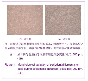
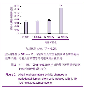
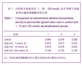
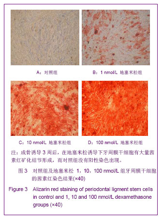
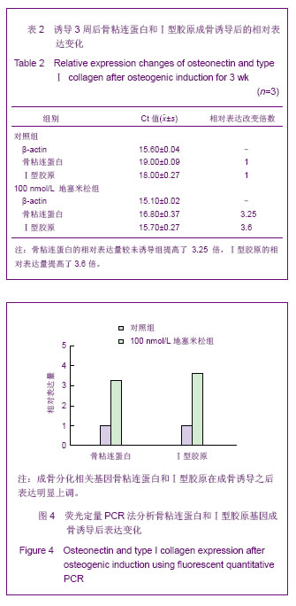
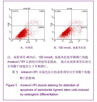
.jpg)