[1] KUSUMBE AP, RAMASAMY SK, ADAMS RH. Coupling of angiogenesis and osteogenesis by a specific vessel subtype in bone. Nature. 2014;507(7492):323-328.
[2] CHEN JY, HENDRIKS M, CHATZIS A, et al. Bone vasculature and bone marrow vascular niches in health and disease. J Bone Miner Res. 2020;35(11):2103-2120.
[3] WANG J, GAO Y, CHENG P, et al. CD31hiEmcnhi vessels support new trabecular bone formation at the frontier growth area in the bone defect repair process. Sci Rep. 2017;7(1):4990.
[4] YIN H, HUANG J, CAO X, et al. Inhibition of src homology 2 domain-containing protein tyrosine phosphatase-2 facilitates CD31hiEndomucinhi blood vessel and bone formation in ovariectomized mice. Cell Physiol Biochem. 2018;50(3):1068-1083.
[5] YIN S, ZHANG W, ZHANG Z, et al. Recent advances in scaffold design and material for vascularized tissue-engineered bone regeneration. Adv Healthc Mater. 2019; 8(10):e1801433.
[6] DAI K, SHEN T, YU Y, et al. Generation of rhBMP-2-induced juvenile ossicles in aged mice. Biomaterials. 2020;258:120284.
[7] CHEN W, XIE G, LU Y, et al. An improved osseointegration of metal implants by pitavastatin loaded multilayer films with osteogenic and angiogenic properties. Biomaterials. 2022;280:121260.
[8] SUN J, JIAO K, NIU L, et al. Intrafibrillar silicified collagen scaffold modulates monocyte to promote cell homing, angiogenesis and bone regeneration. Biomaterials. 2017;113:203-216.
[9] HU J, ZHOU J, WU J, et al. Loganin ameliorates cartilage degeneration and osteoarthritis development in an osteoarthritis mouse model through inhibition of NF-κB activity and pyroptosis in chondrocytes. J Ethnopharmacol. 2020;247:112261.
[10] SHEN Z, CHEN Z, LI Z, et al. Total flavonoids of rhizoma drynariae enhances angiogenic-osteogenic coupling during distraction osteogenesis by promoting type H vessel formation through PDGF-BB/PDGFR-β instead of HIF-1α/VEGF axis. Front Pharmacol. 2020;11:503524.
[11] LI W, ZHOU X, JIANG T, et al. Positive effect of gushukang on type-H vessel and bone formation. Front Cell Dev Biol. 2020;8:265.
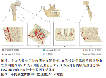
[12] LI H, LIAO L, Hu Y, et al. Identification of type H vessels in mice mandibular condyle. J Dent Res. 2021;100(9):983-992.
[13] YAN Z, WANG X, ZHOU Y, et al. H-type blood vessels participate in alveolar bone remodeling during murine tooth extraction healing. Oral Dis. 2020;26(5):998-1009.
[14] STEFANOWSKI J, LANG A, RAUCH A, et al. Spatial distribution of macrophages during callus formation and maturation reveals close crosstalk between macrophages and newly forming vessels. Front Immunol. 2019;10(2588):2588-2588.
[15] RAMASAMY SK, KUSUMBE AP, Schiller M, et al. Blood flow controls bone vascular function and osteogenesis. Nat Commun. 2016;7:13601-13601.
[16] ROMEO S, ALAWI K, RODRIGUES J, et al. Endothelial proteolytic activity and interaction with non-resorbing osteoclasts mediate bone elongation. Nat Cell Biol. 2019;21(4):430-441.
[17] PENG Y, WU S, LI Y, et al. Type H blood vessels in bone modeling and remodeling. Theranostics. 2020;10(1):426-436.
[18] FANG C, GUO JW, WANG YJ, et al. Diterbutyl phthalate attenuates osteoarthritis in ACLT mice via suppressing ERK/c-fos/NFATc1 pathway, and subsequently inhibiting subchondral osteoclast fusion. Acta Pharmacologica Sinica. 2022;43(5):1299-1310.
[19] LI Y, MU W, XU B, et al. Artesunate, an anti-malaria agent, attenuates experimental osteoarthritis by inhibiting bone resorption and CD31hiEmcnhi vessel formation in subchondral bone. Front Pharmacol. 2019;10:685.
[20] JI B, ZHANG Z, GUO W, et al. Isoliquiritigenin blunts osteoarthritis by inhibition of bone resorption and angiogenesis in subchondral bone. Sci Rep. 2018;8(1):1721.
[21] CUI Z, CRANE J, XIE H, et al. Halofuginone attenuates osteoarthritis by inhibition of TGF-β activity and H-type vessel formation in subchondral bone. Ann Rheum Dis. 2016;75(9):1714-1721.
[22] DING L, GU S, ZHOU B, et al. Ginsenoside compound K enhances fracture healing via promoting osteogenesis and angiogenesis. Front Pharmacol. 2022;13: 855393-855393.
[23] WANG F, QIAN H, KONG L, et al. Accelerated bone regeneration by astragaloside iv through stimulating the coupling of osteogenesis and angiogenesis. Int J Biol Sci. 2021;17(7):1821-1836.
[24] YANG M, LI C, XIAO Y, et al. Ophiopogonin D promotes bone regeneration by stimulating CD31 EMCN vessel formation. Cell Prolif. 2020;53(3):e12784.
[25] LIN X, XU F, ZHANG KW, et al. Acacetin prevents bone loss by disrupting osteoclast formation and promoting type H vessel formation in ovariectomy-induced osteoporosis. Front Cell Dev Biol. 2022;10:796227.
[26] SONG C, CAO J, LEI Y, et al. Nuciferine prevents bone loss by disrupting multinucleated osteoclast formation and promoting type H vessel formation. FASEB J. 2020;34(3):4798-4811.
[27] HUANG J, YIN H, RAO S, et al. Harmine enhances type H vessel formation and prevents bone loss in ovariectomized mice. Theranostics. 2018;8(9):2435-2446.
[28] LIANG S, LING S, DU R, et al. The coupling of reduced type H vessels with unloading-induced bone loss and the protection role of Panax quinquefolium
saponin in the male mice. Bone. 2021;143:115712.
[29] GAO B, LIN X, JING H, et al. Local delivery of tetramethylpyrazine eliminates the senescent phenotype of bone marrow mesenchymal stromal cells and creates an anti-inflammatory and angiogenic environment in aging mice. Aging cell. 2018; 17(3):e12741.
[30] HOOTMAN J, HELMICK C. Projections of US prevalence of arthritis and associated activity limitations. Arthritis Rheum. 2006;54(1):226-229.
[31] LAWRENCE R, FELSON D, HELMICK C, et al. Estimates of the prevalence of arthritis and other rheumatic conditions in the United States. Part II. Arthritis Rheum. 2008;58(1):26-35.
[32] LE GRAVERAND-GASTINEAU M. Disease modifying osteoarthritis drugs: facing development challenges and choosing molecular targets. Current drug targets, 2010;11(5):528-535.
[33] HAWKER G, MIAN S, BEDNIS K, et al. Osteoarthritis year 2010 in review: non-pharmacologic therapy. Osteoarthritis Cartilage. 2011;19(4):366-374.
[34] BERENBAUM F. Osteoarthritis year 2010 in review: non-pharmacological therapies. Osteoarthritis Cartilage. 2011;19(4):361-365.
[35] HUANG J, ZHANG Y, DONG L, et al. Ethnopharmacology, phytochemistry, and pharmacology of Cornus officinalis Sieb. et Zucc. J Ethnopharmacol. 2018;213: 280-301.
[36] WEI CC, YUE LF, YOU FT, et al. Panax notoginseng saponins alleviate osteoporosis and joint destruction in rabbits with antigen‑induced arthritis. Exp Ther Med. 2021;22(5):1302.
[37] 李世杰,马立琼,熊贤梅,等.三七总皂对富血小板血浆促进兔骨缺损愈合的影响[J].中国组织工程研究,2022,26(14):2155-2160.
[38] ZHANG Y, CAI W, HAN G, et al. Panax notoginseng saponins prevent senescence and inhibit apoptosis by regulating the PI3K‑AKT‑mTOR pathway in osteoarthritic chondrocytes. Int J Mol Med. 2020;45(4):1225-1236.
[39] VERMA S, DAS P, KUMAR V L. Chemoprevention by artesunate in a preclinical model of colorectal cancer involves down regulation of β-catenin, suppression of angiogenesis, cellular proliferation and induction of apoptosis. Chem Biol Interact. 2017;278:84-91.
[40] ZHAO C, LIU Q, WANG K. Artesunate attenuates ACLT-induced osteoarthritis by suppressing osteoclastogenesis and aberrant angiogenesis. Biomed Pharmacother. 2017;96:410-416.
[41] FAZZALARI NL. Bone fracture and bone fracture repair. Osteoporos Int. 2011; 22(6):2003-2006.
[42] YANG XD, YANG YY, OUYANG DS, et al. A review of biotransformation and pharmacology of ginsenoside compound K. Fitoterapia. 2015;100:208-220.
[43] YANG N, LIU D, ZHANG X, et al. Effects of ginsenosides on bone remodelling for novel drug applications: a review. Chin Med. 2020;15(1):42.
[44] HUANG Q, GAO B, WANG L, et al. Ophiopogonin D: a new herbal agent against osteoporosis. Bone. 2015;74:18-28.
[45] YANG M, GUO Q, PENG H, et al. Krüppel-like factor 3 inhibition by mutated lncRNA Reg1cp results in human high bone mass syndrome. J Exp Med. 2019; 216(8):1944-1964.
[46] FANG Y, QINGNA L, ZHIHONG T, et al. Effect of total flavonoids from Drynaria rhizome on bone loss in ovariectomized rats. Trop J Pharm Res. 2019;18(6):1285-1289.
[47] ZHANG Y, JIANG J, SHEN H, et al. Total flavonoids from Rhizoma Drynariae (Gusuibu) for treating osteoporotic fractures: implication in clinical practice. Drug Des Devel Ther. 2017;11:1881-1890.
[48] MAO L, XIA L, CHANG J, et al. The synergistic effects of Sr and Si bioactive ions on osteogenesis, osteoclastogenesis and angiogenesis for osteoporotic bone regeneration. Acta Biomater. 2017;61:217-232.
[49] SUN X, GUO Q, WEI W, et al. Current progress on microRNA-based gene delivery in the treatment of osteoporosis and osteoporotic fracture. Int J Endocrinol. 2019;2019:6782653.
[50] DING W, XU C, ZHANG Y, et al. Advances in the understanding of the role of type-H vessels in the pathogenesis of osteoporosis. Arch Osteoporos. 2020;15(1):5.
[51] XIE H, CUI Z, WANG L, et al. PDGF-BB secreted by preosteoclasts induces angiogenesis during coupling with osteogenesis. Nat Med. 2014;20(11):1270-1278.
[52] LI J, LIN X, ZHANG Y, et al. Preparative purification of bioactive compounds from flos chrysanthemi indici and evaluation of its antiosteoporosis effect. Evid Based Complement Alternat Med. 2016;2016:2587201.
[53] KIM SI, KIM YH, KANG BG, et al. Linarin and its aglycone acacetin abrogate actin ring formation and focal contact to bone matrix of bone-resorbing osteoclasts through inhibition of αvβ3 integrin and core-linked CD44. Phytomedicine. 2020; 79:153351.
[54] CHAI S, WAN L, WANG JL, et al. Gushukang inhibits osteocyte apoptosis and enhances BMP-2/Smads signaling pathway in ovariectomized rats. Phytomedicine. 2019;64:153063.
[55] GONG P, ZHANG Z, ZOU Y, et al. Tetramethylpyrazine attenuates blood-brain barrier disruption in ischemia/reperfusion injury through the JAK/STAT signaling pathway. Eur J Pharmacol. 2019;854:289-297.
[56] ZHANG X, DONG H, LIU Y, et al. Tetramethylpyrazine partially relieves hypoxia-caused damage of cardiomyocytes H9c2 by downregulation of miR-449a. J Cell Physiol. 2019. doi: 10.1002/jcp.28151.
[57] GABEL L, LIPHARDT A, HULME P, et al. Pre-flight exercise and bone metabolism predict unloading-induced bone loss due to spaceflight. Br J Sports Med. 2021; 56(4):196-203.
[58] WANG K, WANG Y, HU Z, et al. Bone-targeted lncRNA OGRU alleviates unloading-induced bone loss via miR-320-3p/Hoxa10 axis. Cell Death Dis. 2020;11(5):382.
[59] LI D, LIU M, TAO T Q, et al. Panax quinquefolium saponin attenuates cardiomyocyte apoptosis and opening of the mitochondrial permeability transition pore in a rat model of ischemia/reperfusion. Cell Physiol Biochem. 2014;34(4):1413-1426.
[60] CHEN H, HU B, LV X, et al. Prostaglandin E2 mediates sensory nerve regulation of bone homeostasis. N Nat Commun. 2019;10(1):181.
[61] LI L, LI Q, GUI L, et al. Sequential gastrodin release PU/n-HA composite scaffolds reprogram macrophages for improved osteogenesis and angiogenesis. Bioact Mater. 2022;19:24-37.
[62] ZHAO ZH, MA XL, ZHAO B, et al. Naringin-inlaid silk fibroin/hydroxyapatite scaffold enhances human umbilical cord-derived mesenchymal stem cell-based bone regeneration. Cell Prolif. 2021; 54(7):e13043.
[63] KAO CT, CHIU YC, LEE AK, et al. The synergistic effects of Xu Duan combined Sr-contained calcium silicate/poly-ε-caprolactone scaffolds for the promotion of osteogenesis marker expression and the induction of bone regeneration in osteoporosis. Mater Sci Eng C Mater Biol Appl. 2021;119:111629.
[64] YAN Y, CHEN H, ZHANG H, et al. Vascularized 3D printed scaffolds for promoting bone regeneration. Biomaterials. 2019;190-191:97-110.
[65] 李高志,石菲,张舒,等.血管新生与骨形成耦联、骨骼疾病发生及治疗中H型血管的作用机制研究进展[J].山东医药,2021,61(3):91-94. |
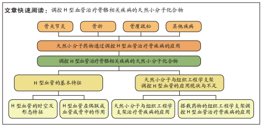
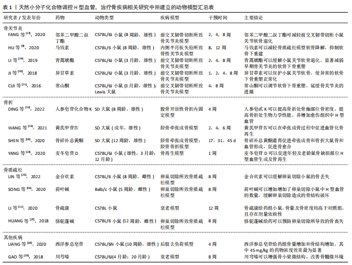
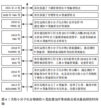
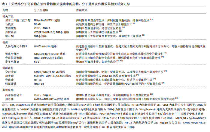 马钱素、异甘草素及常山酮可以通过调节骨关节炎时异常升高的基质金属蛋白酶,来减少H型血管的异常增生,进而减缓关节病变。邻苯二甲酸二叔丁酯与青蒿琥酯可以通过其他通路达到相同效果。在骨折以及牵张成骨的骨愈合过程中,人参皂甙化合物K与黄芪甲苷可以通过 调节β-连环蛋白,促进H型血管生成。骨碎补总黄酮及黄芪甲苷均通过上调血小板衍生生长因子BB,促进H型血管生成。金合欢素、荷叶碱、骆驼蓬碱和骨疏康均可通过不同通路促进H型血管生成,改善骨质疏松。金合欢素与荷叶碱均可抑制多核破骨细胞的成熟和分化,进而减缓骨丧失。此外,在治疗由于衰老以及失重所造成的骨丧失过程中,川穹嗪与西洋参总皂苷,主要通过上调血管内皮生长因子,进而促进H型血管形成与骨再生。在调控H型血管以治疗骨骼相关疾病的研究中,天然小分子化合物显示出良好的前景。搭载天然小分子化合物的组织工程支架在治疗骨相关疾病中,已经得到广泛应用,并且组织工程支架在调控H型血管治疗骨疾病中,也得到了越来越多的关注。所以,将天然小分子化合物与组织工程支架结合到一起,对于调控H型血管治疗骨疾病,是一种具有潜力的新策略。