中国组织工程研究 ›› 2023, Vol. 27 ›› Issue (7): 1055-1061.doi: 10.12307/2023.014

• 组织工程骨材料 tissue-engineered bone • 上一篇    下一篇

杜仲胶夹板固定兔骨折模型的疗效评价

张  伟1,黄致超2,赵锐锋3,梁  欢2,马玉峰1,申艳光4 ,钟红刚5,陈兆军1,张继川3,陈卫衡1   

  1. 北京中医药大学第三附属医院,1骨伤中心,4放射科,北京中医药大学,北京市  100029;2北京中医药大学研究生院,北京市  100029;3北京化工大学材料科学与工程学院,先进弹性体材料研究中心,北京市  100029;5中国中医科学院骨伤研究所,北京市  100700
  • 收稿日期:2021-09-22 接受日期:2021-12-15 出版日期:2023-03-08 发布日期:2022-07-18
  • 通讯作者: 陈卫衡,博士,主任医师,北京中医药大学第三附属医院骨伤中心,北京市 100029
  • 作者简介:张伟,女,1988年生,河北省邯郸市人,汉族,博士,助理研究员,主要从事中医药治疗骨伤病的应用基础研究。
  • 基金资助:
    北京中医药大学新教师启动项目(2020-JYB-XJSJJ-058),项目负责人:张伟;北京中医药大学高层次人才启动项目支持(2021-XJ-KYQD-001),项目负责人:陈卫衡

Efficacy of gutta-percha splint on a rabbit fracture model

Zhang Wei1, Huang Zhichao2, Zhao Ruifeng3, Liang Huan2, Ma Yufeng1, Shen Yanguang4, Zhong Honggang5, Chen Zhaojun1, Zhang Jichuan3, Chen Weiheng1   

  1. 1Traumatology & Orthopedics Center, Third Affiliated Hospital of Beijing University of Chinese Medicine, Beijing 100029; 2Graduate School, Beijing University of Chinese Medicine, Beijing 100029; 3College of Materials Science and Engineering, Beijing University of Chemical Technology, Beijing 100029; 4Imaging Center, Third Affiliated Hospital of Beijing University of Chinese Medicine, Beijing 100029; 5Institute of Orthopedics, China Academy of Traditional Chinese Medicine, Beijing 100700
  • Received:2021-09-22 Accepted:2021-12-15 Online:2023-03-08 Published:2022-07-18
  • Contact: Chen Weiheng, MD, Chief physician, Traumatology & Orthopedics Center, Third Affiliated Hospital of Beijing University of Chinese Medicine, Beijing 100029, China
  • About author:Zhang Wei, MD, Assistant researcher, Traumatology & Orthopedics Center, Third Affiliated Hospital of Beijing University of Chinese Medicine, Beijing 100029, China
  • Supported by:
    New Teacher Start-Up Project of Beijing University of Chinese Medicine, No. 2020-JYB-XJSJJ-058 (to ZW); Key Research Project of Beijing University of Chinese Medicine No. 2021-XJ-KYQD-001 ( to CWH)

摘要:

文题释义:
杜仲胶:是含于杜仲树的丝状物质,为一种天然高分子材料,具有橡塑二重特性,此力学性能有望为骨折愈合的弹性固定提供新材料。
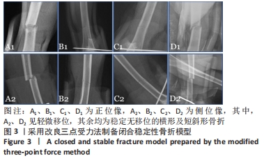
闭合骨折模型:主要利用三点受力原理模拟长骨干骨折时的受力情况,借助带有重物的钝性铡刀从高处落下,折断胫骨或股骨则造模成功,具有可重复性高、与临床实际情况相似度高的优势,被广泛应用于骨折后愈合过程的研究。

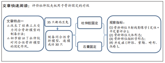
背景:课题组成员前期研究发现杜仲胶的屈服强度较低,呈现硬而韧的力学性能,在此基础上研发了根据体型自由塑形的运动护具,并初步证实此性能可降低外界冲击力对运动员的伤害。
目的:评价杜仲胶夹板用于骨折固定的疗效。
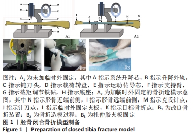
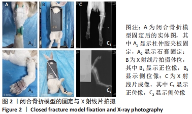
方法:取35只成年雄性新西兰兔,采用改良三点受力法制备建立左侧胫骨闭合性骨折模型。将造模成功的30只兔随机分为2组,分别采用杜仲胶夹板和石膏进行外固定,每组15只。于固定后2 h、4周、6周进行X射线片检测,观察固定后骨折断端愈合情况;固定6周后,分别进行胫骨标本大体与组织学形态观察。
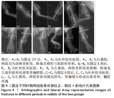
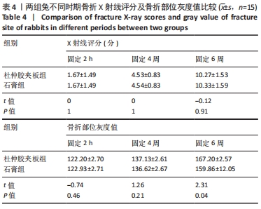
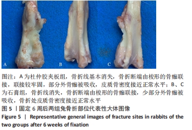
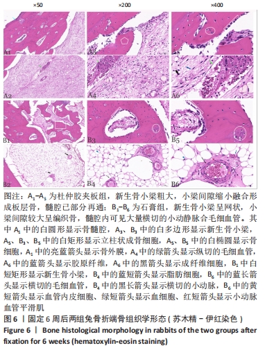
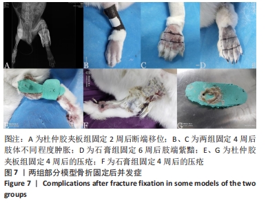
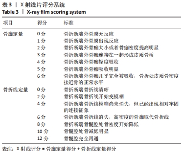
结果与结论:①X射线片:固定2 h后,两组均可见清晰骨折线,均未见明显骨膜反应;固定4周后,两组骨折线开始变模糊,断端见大面积梭形低密度骨痂阴影;固定6周后,两组骨折线接近消失,骨痂连接在一起形成皮质骨桥,骨髓腔部分已通、部分接近再通,固定6周后,杜仲胶夹板组骨折部位灰度值高于石膏组(P < 0.05);②大体观察:杜仲胶夹板组除3只出现畸形愈合外,其余兔骨折线基本消失,部分外骨痂被吸收,皮质骨密度接近正常水平;石膏组除4只出现畸形愈合外,其余兔骨折线消失,少部分外骨痂被吸收,皮质骨密度接近正常水平;③组织学形态:杜仲胶夹板组可见粗大的新生骨小梁,小梁间隙缩小融合形成板层骨,髓腔已部分再通;石膏组新生骨小梁呈网状,形成编织骨,髓腔内可见大量横切的小动静脉和毛细血管;④并发症:固定6周内,杜仲胶夹板组肢端肿胀、紫黯、移位及畸形愈合、局部压疮数量存在少于石膏组的趋势,但统计学分析比较差异均无显著性意义(P > 0.05);⑤结果表明,杜仲胶夹板对胫骨闭合性骨折兔模型可起到与石膏类似的固定作用。

https://orcid.org//0000-0001-8692-0913(张伟)

中国组织工程研究杂志出版内容重点:生物材料;骨生物材料口腔生物材料纳米材料缓释材料材料相容性组织工程

关键词: 杜仲胶, 石膏, 夹板, 闭合性骨折, 组织病理学, 并发症, 新西兰兔

Abstract: BACKGROUND:  Gutta-percha has low yield strength and shows hard and tough mechanical properties. On this basis, a sports protective gear that can be freely shaped according to the body shape has been developed, and it has been preliminarily confirmed that this performance can reduce the damage to athletes caused by external impact forces.
OBJECTIVE: To evaluate the effect of gutta-percha splint on fracture fixation.
METHODS: Thirty-five adult male New Zealand rabbits were used to establish closed fracture models of the left tibia by the modified three-point stress method. Totally 30 successful models were randomly divided into two groups (n=15) and fixed with gutta-percha splint and gypsum, respectively. The effects of the two external fixation methods were compared by X-ray imaging and fracture healing was observed at 2 hours, 4 and 6 weeks after fixation. After 6 weeks of fixation, the tibia specimens were observed for gross and histological morphology.
RESULTS AND CONCLUSION: (1) X-ray films: Clear fracture line was seen 2 hours after fixation, without obvious periosteal reaction. After 4 weeks, the fracture line became blurred, and a large area of spindle low-density callus shadow was seen at the broken end. After 6 weeks, the fracture line nearly disappeared; the callus connected together to form a cortical bone bridge, and the bone marrow cavity was partially canalized, and partially close to recanalization. After 6 weeks, gray value at the fracture site was higher in the gutta-percha splint group than that in the gypsum group (P < 0.05). (2) General observation: In the gutta-percha splint group, except for three rabbits with malunion, the fracture line of the rest of the rabbits basically disappeared; part of the outer callus was absorbed, and the cortical bone density was close to the normal level. In the gypsum group, except for four rabbits with malunion, the fracture line of the rest of the rabbits disappeared, a small part of the outer callus was absorbed, and the cortical bone density was close to the normal level. (3) Histological morphology: In the gutta-percha splint group, new bone trabeculae were thick; the trabecular space was reduced and fused to form lamellar bone, and the medullary cavity had been partially recanalized. In the gypsum group, new bone trabeculae were reticular, forming a woven bone, and a large number of transected small arteries, veins and capillaries could be seen in the medullary cavity. (4) Complications: Within 6 weeks of fixation, the extremity swelling, purple darkening, displacement and malunion, and the number of local pressure ulcers in the gutta-percha splint group tended to be less than those in the gypsum group, but there was no significant difference in statistical analysis (P > 0.05). (5) The results showed that gutta-percha splint could play a similar fixation effect to plaster on the rabbit model of closed tibial fracture. 

Key words: gutta-percha, gypsum, splint, closed fracture, histopathology, complication, New Zealand rabbit

中图分类号: