中国组织工程研究 ›› 2023, Vol. 27 ›› Issue (18): 2897-2902.doi: 10.12307/2023.348

• 骨与关节图像与影像 bone and joint imaging • 上一篇    下一篇

椎体后方结构及对突出椎间盘体积测量的影响

牛策号,张春霖,严  旭,付  苏,冯  阳,朱安迪   

  1. 郑州大学第一附属医院骨科,河南省郑州市   450000
  • 收稿日期:2022-01-25 接受日期:2022-05-17 出版日期:2023-06-28 发布日期:2022-09-19
  • 通讯作者: 张春霖,博士,主任医师,教授,郑州大学第一附属医院骨科,河南省郑州市 450000
  • 作者简介:牛策号,男,1989年生,河北省邯郸市人,汉族,郑州大学第一附属医院在职硕士,河北省邯郸市武安市第一人民医院骨科主治医师,主要从事微创脊柱外科研究。

Posterior vertebral structure and its influence on measurement of disc herniation volume

Niu Cehao, Zhang Chunlin, Yan Xu, Fu Su, Feng Yang, Zhu Andi   

  1. Department of Orthopedics, First Affiliated Hospital of Zhengzhou University, Zhengzhou 450000, Henan Province, China
  • Received:2022-01-25 Accepted:2022-05-17 Online:2023-06-28 Published:2022-09-19
  • Contact: Zhang Chunlin, MD, Chief physician, Professor, Department of Orthopedics, First Affiliated Hospital of Zhengzhou University, Zhengzhou 450000, Henan Province, China
  • About author:Niu Cehao, Master candidate, Attending physician, Department of Orthopedics, First Affiliated Hospital of Zhengzhou University, Zhengzhou 450000, Henan Province, China

摘要:

文题释义:
椎体后方结构:是椎管内椎体和前侧硬膜之间的结构,前边界为椎体后缘和椎间盘后缘,后边界为脊髓/脑脊液,外侧为椎间孔和椎弓根,上端和底端分别为枕骨大孔和骶骨裂孔。其中的组织主要包括:后纵韧带、疏松蜂窝状结缔组织、椎管内静脉丛、脂肪组织、硬膜外韧带、淋巴管等,功能集中于为脊柱提供力学支持,且具有血供、营养、连接和缓冲保护作用。
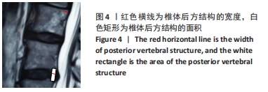
三维体积测量法:是利用PACS软件在MRI矢状位T2图像上,以上位椎体后下缘及下位椎体后上缘连线为内边界,突出物边缘作为外边界,得到当前层面突出腰椎间盘的面积,逐一测量每个层面的面积,利用以下公式计算突出腰椎间盘体积:突出腰椎间盘体积=(层间距+层厚)×∑每一层突出腰椎间盘面积。

背景:突出椎间盘体积测量是定量评估椎间盘突出程度的重要数据,以往在对内窥镜下微创颈椎管成形术后突出颈椎间盘体积的观察中发现,定量测量不能证实突出的颈椎间盘被完全吸收,可能与椎体后方一些不能吸收回纳的组织被包容在突出的椎间盘组织中一并被测量有关。
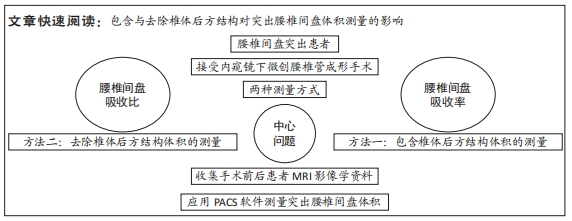
目的:对比分析包含与去除椎体后方结构对突出腰椎间盘体积测量的影响,并初步观察内镜下微创腰椎管成形术后突出椎间盘体积的早期变化。
方法:选择郑州大学第一附属医院收治的腰椎间盘突出患者98例(涉及191个椎间盘),收集患者内镜下微创腰椎管成形术前与术后1周的腰椎MRI影像资料,均应用PACS软件测量突出椎间盘体积,分2种测量方法:一组包含椎体后方结构,另一组去除椎体后方结构。比较两种测量方式下,手术前后突出腰椎间盘的缩小率、缩小比、不变比、增大比、增大率,以及术后1周的2种测量方法下的突出腰椎间盘的吸收比、吸收率。
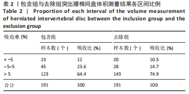
结果与结论:①包含组术后突出腰椎间盘体积缩小的有123个,缩小比64.4%,最大缩小率34.17%;体积不变的有45个,不变比23.6%;体积增大的有23个,增大比12%,最大增大率59.9%。去除组术后突出腰椎间盘体积缩小的有143个,缩小比74.9%,最大缩小率51.47%;体积不变的有28个,不变比14.7%;体积增大的有20个,增大比10.5%,最大增大率60.26%。②包含组术后突出腰椎间盘的吸收比、吸收率均低于去除组(P < 0.01)。③结果显示,去除椎体后方结构进行突出腰椎间盘体积测量的方法更为接近实际状态,精准度更高;内镜下微创腰椎管成形术后早期即能广泛“诱导”突出的腰椎间盘发生自然吸收。

https://orcid.org/0000-0002-3848-2266 (牛策号);https://orcid.org/0000-001-9565-4485 (张春霖)

中国组织工程研究杂志出版内容重点:人工关节;骨植入物;脊柱;骨折;内固定;数字化骨科;组织工程

关键词: 内镜下微创腰椎管成形术, 突出腰椎间盘, 自然吸收, 突出腰椎间盘体积, 椎体后方结构, MRI

Abstract: BACKGROUND: Volume of disc herniation is an important measurement index for quantitative evaluation of disc herniation degree. Previously, the volume of cervical disc herniation that was completely absorbed after cervical microendoscopic laminoplasty was found unable to be confirmed by volume quantitative measurement. The reason was related to the fact that some tissues posterior to the vertebral body that could not be absorbed were included during measurement.  
OBJECTIVE: To compare and analyze the effect of inclusion and removal of the posterior vertebral structure on the volume measurement of herniated lumbar intervertebral discs, and to preliminarily observe the early changes of the volume of herniated intervertebral discs after lumbar microendoscopic laminoplasty.
METHODS:  Totally 98 patients with lumbar disc herniation (191 intervertebral discs) in First Affiliated Hospital of Zhengzhou University were enrolled. Lumbar spine MRI images of patients before and 1 week after lumbar microendoscopic laminoplasty were collected. The volumes were measured using PACS software. There were two measurement methods: one group included the posterior vertebral structure and the other group removed the posterior vertebral structure. The reduction rate, reduction ratio, unchanged ratio, enlargement ratio, and enlargement rate of the herniated lumbar intervertebral discs before and after the operation under the two measurement methods, and the absorption ratio and absorption rate of the herniated lumbar intervertebral disc under two measurement methods at 1 week after operation were compared.  
RESULTS AND CONCLUSION: (1) In the inclusion group, 123 disc herniations with reduced volume after lumbar microendoscopic laminoplasty (reduction ratio of 64.4%, maximum reduction rate of 34.17%); 45 disc herniations were unchanged in volume, ratio of 23.6%; 23 disc herniations showed increase in volume, increase ratio of 12%, and maximum increase rate of 59.9%. In the exclusion group, 143 disc herniations were reduced in volume after lumbar microendoscopic laminoplasty (reduction ratio of 74.9%, maximum reduction rate of 51.47%), 28 disc herniations were shown unchanged in volume, constant ratio of 14.7%, and 20 disc herniations increased in volume, increase ratio of 10.5%, maximum increase rate of 60.26%. (2) The absorption ratio and absorption rate of herniated lumbar intervertebral disc were lower in the inclusion group than that in the exclusion group (P < 0.01). (3) It is concluded that the measurement method of disc herniation volume by exclusion of posterior vertebral structure is closer to the actual state with a high accuracy. Lumbar microendoscopic laminoplasty can induce the herniated lumbar disc to spontaneously absorb in the early stage postoperatively.

Key words: lumbar microendoscopic laminoplasty, herniated lumbar intervertebral disc, natural absorption, herniated lumbar disc volume, posterior vertebral structure, MRI

中图分类号: