[1] HOLMES D. Spinal-cord injury: spurring regrowth. Nature. 2017;552 (7684):S49.
[2] AHUJA CS, WILSON JR, NORI S, et al. Traumatic spinal cord injury. Nat Rev Dis Primers. 2017;3:17018.
[3] LOANE DJ, FADEN AI. Neuroprotection for traumatic brain injury: translational challenges and emerging therapeutic strategies. Trends Pharmacol Sci. 2010;31(12):596-604.
[4] DORRIER CE, JONES HE, PINTARIĆ L, et al. Emerging roles for CNS fibroblasts in health, injury and disease. Nat Rev Neurosci. 2022;23(1): 23-34.
[5] BRESLIN K, AGRAWAL D. The use of methylprednisolone in acute spinal cord injury: a review of the evidence, controversies, and recommendations. Pediatr Emerg Care. 2012;28(11):1238-1245; quiz 1246-1248.
[6] HURLBERT RJ. Methylprednisolone for the treatment of acute spinal cord injury: point. Neurosurgery. 2014;61 Suppl 1:32-35.
[7] GONCALVES MB, MALMQVIST T, CLARKE E, et al. Neuronal RARβ Signaling Modulates PTEN Activity Directly in Neurons and via Exosome Transfer in Astrocytes to Prevent Glial Scar Formation and Induce Spinal Cord Regeneration. J Neurosci. 2015;35(47):15731-15745.
[8] GONCALVES MB, WU Y, CLARKE E, et al. Regulation of Myelination by Exosome Associated Retinoic Acid Release from NG2-Positive Cells. J Neurosci. 2019;39(16):3013-3027.
[9] GONCALVES MB, WU Y, TRIGO D, et al. Retinoic acid synthesis by NG2 expressing cells promotes a permissive environment for axonal outgrowth. Neurobiol Dis. 2018;111:70-79.
[10] PAN D, LI Y, YANG F, et al. Increasing toll-like receptor 2 on astrocytes induced by Schwann cell-derived exosomes promotes recovery by inhibiting CSPGs deposition after spinal cord injury. J Neuroinflammation. 2021;18(1):172.
[11] PAN D, ZHU S, ZHANG W, et al. Autophagy induced by Schwann cell-derived exosomes promotes recovery after spinal cord injury in rats. Biotechnol Lett. 2022;44(1):129-142.
[12] WEBBER C, ZOCHODNE D. The nerve regenerative microenvironment: early behavior and partnership of axons and Schwann cells. Exp Neurol. 2010;223(1):51-59.
[13] AHUJA CS, SCHROEDER GD, VACCARO AR, et al. Spinal Cord Injury-What Are the Controversies? J Orthop Trauma. 2017;31 Suppl 4:S7-S13.
[14] DE RIVERO VACCARI JP, DIETRICH WD, KEANE RW. Therapeutics targeting the inflammasome after central nervous system injury. Transl Res. 2016;167(1):35-45.
[15] GAO ZS, ZHANG CJ, XIA N, et al. Berberine-loaded M2 macrophage-derived exosomes for spinal cord injury therapy. Acta Biomater. 2021; 126:211-223.
[16] KIM GU, SUNG SE, KANG KK, et al. Therapeutic Potential of Mesenchymal Stem Cells (MSCs) and MSC-Derived Extracellular Vesicles for the Treatment of Spinal Cord Injury. Int J Mol Sci. 2021;22(24):13672.
[17] CARUSO BAVISOTTO C, SCALIA F, MARINO GAMMAZZA A, et al. Extracellular Vesicle-Mediated Cell⁻Cell Communication in the Nervous System: Focus on Neurological Diseases. Int J Mol Sci. 2019;20(2):434.
[18] CIREGIA F, URBANI A, PALMISANO G. Extracellular Vesicles in Brain Tumors and Neurodegenerative Diseases. Front Mol Neurosci. 2017;10:276.
[19] HOLM MM, KAISER J, SCHWAB ME. Extracellular Vesicles: Multimodal Envoys in Neural Maintenance and Repair. Trends Neurosci. 2018;41(6): 360-372.
[20] SAEEDI S, ISRAEL S, NAGY C, et al. The emerging role of exosomes in mental disorders. Transl Psychiatry. 2019;9(1):122.
[21] YATES AG, ANTHONY DC, RUITENBERG MJ, et al. Systemic Immune Response to Traumatic CNS Injuries-Are Extracellular Vesicles the Missing Link? Front Immunol. 2019;10:2723.
[22] DUTTA D, KHAN N, WU J, et al. Extracellular Vesicles as an Emerging Frontier in Spinal Cord Injury Pathobiology and Therapy. Trends Neurosci. 2021;44(6):492-506.
[23] ZHANG B, YEO RW, TAN KH, et al. Focus on Extracellular Vesicles: Therapeutic Potential of Stem Cell-Derived Extracellular Vesicles. Int J Mol Sci. 2016;17(2):174.
[24] ZHOU Y, WEN LL, LI YF, et al. Exosomes derived from bone marrow mesenchymal stem cells protect the injured spinal cord by inhibiting pericyte pyroptosis. Neural Regen Res. 2022;17(1):194-202.
[25] LOPEZ-VERRILLI MA, COURT FA. Transfer of vesicles from schwann cells to axons: a novel mechanism of communication in the peripheral nervous system. Front Physiol. 2012;3:205.
[26] SERINI G, BUSSOLINO F. Common cues in vascular and axon guidance. Physiology (Bethesda). 2004;19:348-354.
[27] ADAMS RH, EICHMANN A. Axon guidance molecules in vascular patterning. Cold Spring Harb Perspect Biol. 2010;2(5):a001875.
[28] HIMMELS P, PAREDES I, ADLER H, et al. Motor neurons control blood vessel patterning in the developing spinal cord. Nat Commun. 2017;8: 14583.
[29] SILVA NA, SOUSA N, REIS RL, et al. From basics to clinical: a comprehensive review on spinal cord injury. Prog Neurobiol. 2014;114:25-57.
[30] TRAN AP, WARREN PM, SILVER J. The Biology of Regeneration Failure and Success After Spinal Cord Injury. Physiol Rev. 2018;98(2):881-917.
[31] CARMELIET P. Mechanisms of angiogenesis and arteriogenesis. Nat Med. 2000;6(4):389-395.
[32] CASELLA GT, MARCILLO A, BUNGE MB, et al. New vascular tissue rapidly replaces neural parenchyma and vessels destroyed by a contusion injury to the rat spinal cord. Exp Neurol. 2002;173(1):63-76.
[33] FASSBENDER JM, WHITTEMORE SR, HAGG T. Targeting microvasculature for neuroprotection after SCI. Neurotherapeutics. 2011;8(2):240-251.
[34] LOSEY P, YOUNG C, KRIMHOLTZ E, et al. The role of hemorrhage following spinal-cord injury. Brain Res. 2014;1569:9-18.
[35] LOY DN, CRAWFORD CH, DARNALL JB, et al. Temporal progression of angiogenesis and basal lamina deposition after contusive spinal cord injury in the adult rat. J Comp Neurol. 2002;445(4):308-324.
[36] NG MT, STAMMERS AT, KWON BK. Vascular disruption and the role of angiogenic proteins after spinal cord injury. Transl Stroke Res. 2011; 2(4):474-491.
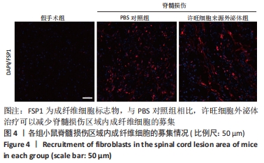
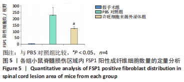
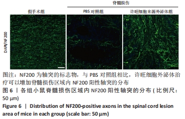
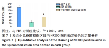
|