[1] LIU M, LV Y. Reconstructing Bone with Natural Bone Graft: A Review of In Vivo Studies in Bone Defect Animal Model.Nanomaterials (Basel). 2018;8(12).pii:E999.doi: 10.3390/nano8120999.
[2] BALDWIN P, LI DJ, AUSTON DA, et al.Autograft, Allograft, and Bone Graft Substitutes: Clinical Evidence and Indications for Use in the Setting of Orthopaedic Trauma Surgery.J Orthop Trauma.2019;33(4):203-213.
[3] GRIGORIAN SHAMAGIAN L, MADONNA R, TAYLOR D, et al. Perspectives on Directions and Priorities for Future Preclinical Studies in Regenerative Medicine.Circ Res.2019;124(6):938-951.
[4] ORYAN A, ALIDADI S, MOSHIRI A, et al. Platelet-rich plasma for bone healing and regeneration. Expert Opin Biol Ther.2016;16(2):213-232.
[5] WANG SJ, JIANG D, ZHANG ZZ, et al. Biomimetic Nanosilica-Collagen Scaffolds for In Situ Bone Regeneration: Toward a Cell-Free, One-Step Surgery.Adv Mater.2019;31(49):e1904341.
[6] FILARDO G, ROFFI A, FEY T, et al. Vegetable hierarchical structures as template for bone regeneration: New bio-ceramization process for the development of a bone scaffold applied to an experimental sheep model.J Biomed Mater Res B Appl Biomater.2020;108(3):600-611.
[7] LI JT, XIONG SL, DING LH, et al. The mechanism research of non-Smad dependent TAK1 signaling pathway in the treatment of bone defects by recombination BMP-2-loaded hollow hydroxyapatite microspheres/chitosan composite.J Mater Sci Mater Med.2019;30(12):130.
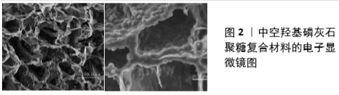
[8] YAO AH, LI XD, XIONG L, et al. Hollow hydroxyapatite microspheres/chitosan composite as a sustained delivery vehicle for rhBMP-2 in the treatment of bone defects.J Mater Sci Mater Med.2015;26(1):5336.
[9] XIONG L, ZENG JH, YAO AH, et al. BMP2-loaded hollow hydroxyapatite microspheres exhibit enhanced osteoinduction and osteogenicity in large bone defects.Int J Nanomedicine 2015;10:517-526.
[10] XIAO W, BAL BS, RAHAMAN MN. Preparation of resorbable carbonate-substituted hollow hydroxyapatite microspheres and their evaluation in osseous defects in vivo.Mater Sci Eng C Mater Biol Appl.2016;60: 324-332.
[11] TANAKA T, TAKAI Y, NAGASE A, et al. Protein adsorption characteristics of nanoparticle-assembled hollow microspheres of hydroxyapatite and their composites with PLLA microporous membranes.Heliyon. 2019;5(4):e01490.
[12] ZENG JH, XIONG SL, DING LH, et al. Study of bone repair mediated by recombination BMP-2/ recombination CXC chemokine Ligand-13-loaded hollow hydroxyapatite microspheres/chitosan composite.Life Sci.2019;234:116743.
[13] Zeng JH, Liu SW, Xiong L, et al. Scaffolds for the repair of bone defects in clinical studies: a systematic review.J Orthop Surg Res. 2018; 13(1):33.
[14] LEE JH, KIM HW. Emerging properties of hydrogels in tissue engineering.J Tissue Eng. 2018;9:2041731418768285.
[15] WANG K, WANG X, HAN C, et al. From Micro to Macro: The Hierarchical Design in a Micropatterned Scaffold for Cell Assembling and Transplantation. Adv Mater.2017;29(2).doi: 10.1002/adma.201604600.
[16] ZHANG C, YUAN HH, LIU HH, et al. Well-aligned chitosan-based ultrafine fibers committed teno-lineage differentiation of human induced pluripotent stem cells for Achilles tendon regeneration. Biomaterials.2015;53:716-730.
[17] HOLTHAUS MG, STOLLE J, TRECCANI L, et al. Orientation of human osteoblasts on hydroxyapatite-based microchannels.Acta Biomater. 2012;8(1):394-403.
[18] RIVELINE D, ZAMIR E, BALABAN NQ, et al. Focal contacts as mechanosensors: externally applied local mechanical force induces growth of focal contacts by an mDia1-dependent and ROCK-independent mechanism.J Cell Biol.2001;153(6):1175-1186.
[19] WEINER S, WAGNER D. The Material Bone: Structure-Mechanical Function Relations.Annu Rev Mater Res.2003;28:271-298.
[20] LIU Y, LUO D, WANG T. Hierarchical Structures of Bone and Bioinspired Bone Tissue Engineering.Small.2016;12(34):4611-4632.
[21] REZNIKOV N, SHAHAR R, WEINER S. Three-dimensional structure of human lamellar bone: The presence of two different materials and new insights into the hierarchical organization. Bone.2014;59:93-104.
[22] RHO JY, KUHN L, ZIOUPOS P. Mechanical Properties and the Hierarchical Structure of Bone. Med Eng Phys.1998;20(2):92-102.
[23] WON JE, LEE YS, PARK JH, et al. Hierarchical microchanneled scaffolds modulate multiple tissue-regenerative processes of immune-responses, angiogenesis, and stem cell homing.Biomaterials. 2020; 227:119548.
[24] YANG H, ZHANG D, PENG WK, et al. Facile synthesis of copper doping hierarchical hollow porous hydroxyapatite beads by rapid gelling strategy.Mater Sci Eng C Mater Biol Appl. 2020;109:110531.
[25] SU N, GAO PL, WANG K, et al. Fibrous scaffolds potentiate the paracrine function of mesenchymal stem cells: A new dimension in cell-material interaction.Biomaterials. 2017;141:74-85.
[26] GNECCHI M, ZHANG ZP, NI AG, et al. Paracrine mechanisms in adult stem cell signaling and therapy.Circ Res.2008;103(11):1204-1219.
[27] SINGER NG, CAPLAN AI. Mesenchymal Stem Cells: Mechanisms of Inflammation.Annu Rev Pathol.2011;6:457-478.
[28] THEVENOT PT, NAIR AM, SHEN J, et al. The effect of incorporation of SDF-1alpha into PLGA scaffolds on stem cell recruitment and the inflammatory response.Biomaterials. 2010;31(14):3997-4008.
[29] OGLE ME, SEGAR CE, SRIDHAR S, et al. Monocytes and macrophages in tissue repair: Implications for immunoregenerative biomaterial design.Exp Biol Med (Maywood). 2016;241(10):1084-1097.
[30] PEI X, MA L, ZHANG B, et al. Creating hierarchical porosity hydroxyapatite scaffolds with osteoinduction by three-dimensional printing and microwave sintering. Biofabrication.2017;9(4):045008.
[31] WANG XJ, LOU T, ZHAO WH, et al. The effect of fiber size and pore size on cell proliferation and infiltration in PLLA scaffolds on bone tissue engineering.J Biomater Appl.2016;30(10):1545-1551.
[32] BILLIET T, GEVAERT E, DE SCHRYVER T, et al. The 3D printing of gelatin methacrylamide cell-laden tissue-engineered constructs with high cell viability.Biomaterials. 2014;35(1):49-62.
[33] HONG Y, HUBER A, AKANARI K, et al. Mechanical properties and in vivo behavior of a biodegradable synthetic polymer microfiber-extracellular matrix hydrogel biohybrid scaffold.Biomaterials. 2011;32(13):3387-3394.
[34] LIU X, WU S, YEUNG KWK, et al. Relationship between osseointegration and superelastic biomechanics in porous NiTi scaffolds. Biomaterials. 2011;32(2):330-338.
[35] XU C, SU P, CHEN X, et al. Biocompatibility and osteogenesis of biomimetic Bioglass-Collagen-Phosphatidylserine composite scaffolds for bone tissue engineering. Biomaterials.2011;32(4):1051-1058.
[36] BARBIERI D, YUAN H, LUO X, et al. Influence of polymer molecular weight in osteoinductive composites for bone tissue regeneration.Acta Biomater.2013;9(12):9401-9413.
[37] GROEN N, YUAN H, HEBELS DGAJ, et al. Linking the Transcriptional Landscape of Bone Induction to Biomaterial Design Parameters.Adv Mater.2017;29(10).doi:10.1002/adma.201603259
[38] LEE VK, DAI GH. Printing of Three-Dimensional Tissue Analogs for Regenerative Medicine. Ann Biomed Eng.2017;45(1):115-131.
[39] ZHOU C, YE X, FAN Y, et al. Biomimetic fabrication of a three-level hierarchical calcium phosphate/collagen/hydroxyapatite scaffold for bone tissue engineering. Biofabrication.2014;6(3):035013.
[40] Li X, Ding J, Wang J, et al.Biomimetic biphasic scaffolds for osteochondral defect repair. Regen Biomater.2015;2(3):221-228.
[41] BUSCHMANN I, SCHAPER W. Arteriogenesis Versus Angiogenesis: Two Mechanisms of Vessel Growth.News Physiol Sci.1999;14:121-125.
[42] BUSCHMANN I, SCHAPER W. The pathophysiology of the collateral circulation (arteriogenesis).J Pathol.2000;190(3):338-342.
[43] PAN HH, XIE YT, ZHANG ZQ, et al. Immunomodulation effect of a hierarchical macropore/nanosurface on osteogenesis and angiogenesis.Biomed Mater.2017;12(4):045006.
[44] MADDEN LR, MORTISEN DJ, SUSSMAN EM, et al. Proangiogenic scaffolds as functional templates for cardiac tissue engineering.Proc Natl Acad Sci U S A.2010;107(34):15211-15216.
[45] YU T, LIU Q, JIANG T, et al. Channeled β-TCP Scaffolds Promoted Vascularization and Bone Augmentation in Mandible of Beagle Dogs.Adv Funct Mater.2016;26(37):6719-6727.
[46] RATNER BD. Reducing capsular thickness and enhancing angiogenesis around implant drug release systems.J Control Release. 2002;78 (1-3): 211-218.
[47] TIAN T, ZHANG T, LIN Y, et al. Vascularization in Craniofacial Bone Tissue Engineering.J Dent Res.2018;97(9):969-976.
[48] DARNELL M, YOUNG S, GU L, et al. Substrate Stress-Relaxation Regulates Scaffold Remodeling and Bone Formation In Vivo.Adv Healthc Mater. 2017;6(1):10.1002/adhm.201601185.
[49] YU T, DONG C, SHEN ZH, et al. Vascularization of plastic calcium phosphate cement in vivo induced by in-situ-generated hollow channels. Mater Sci Eng C Mater Biol Appl. 2016;68:153-162.
[50] ARINZEH TL, PETER SJ, ARCHAMBAULT MP, et al. Allogeneic mesenchymal stem cells regenerate bone in a critical-sized canine segmental defect.J Bone Joint Surg Am. 2003;85(10):1927-1935.
[51] XING HL, WANG X, XIAO G, et al. Hierarchical assembly of nanostructured coating for siRNA-based dual therapy of bone regeneration and revascularization.Biomaterials. 2020;235:119784.
[52] JANG CH, LEE J, KIM G. Synergistic effect of alginate/BMP-2/Umbilical cord serum-coated on 3D-printed PCL biocomposite for mastoid obliteration model.J Ind Eng Chem.2019;72:432-441.
|