中国组织工程研究 ›› 2019, Vol. 23 ›› Issue (24): 3829-3833.doi: 10.3969/j.issn.2095-4344.1232

• 骨与关节生物力学 bone and joint biomechanics • 上一篇    下一篇

股骨近端几何结构结合骨密度预测帕金森病髋部骨折的风险

王武华1,刘旭东2,胡  凌3
  

  1. 1南昌大学抚州医学院影像教研室,江西省抚州市  344000;2南昌大学抚州医学院,江西省抚州市  344000;3抚州市第一人民医院影像二科,江西省抚州市  344000
  • 出版日期:2019-08-28 发布日期:2019-08-28
  • 通讯作者: 刘旭东,硕士,主任医师,副教授,南昌大学抚州医学院,江西省抚州市 344000
  • 作者简介:王武华,男,1987年生,江西省抚州市人,汉族,2015年福建医科大学毕业,硕士,医师,主要从事神经系统疾病与影像方面的研究。
  • 基金资助:
    抚州市社会发展指导性科技计划项目[抚科计字2016(10-4)]

Prediction of hip fracture in Parkinson’s disease with the combination of geometric structure of proximal femur and bone mineral density   

Wang Wuhua1, Liu Xudong2, Hu Ling3
  

  1. 1Department of Radiology, Fuzhou Medical College of Nanchang University, Fuzhou 344000, Jiangxi Province, China; 2Fuzhou Medical College of Nanchang University, Fuzhou 344000, Jiangxi Province, China; 3Second Department of Radiology, the First People’s Hospital of Fuzhou, Fuzhou 344000, Jiangxi Province, China
  • Online:2019-08-28 Published:2019-08-28
  • Contact: Liu Xudong, Master, Chief physician, Associate professor, Fuzhou Medical College of Nanchang University, Fuzhou 344000, Jiangxi Province, China
  • About author:Wang Wuhua, Master, Physician, Department of Radiology, Fuzhou Medical College of Nanchang University, Fuzhou 344000, Jiangxi Province, China
  • Supported by:
    the Social Development Guiding Science and Technology Program of Fuzhou, No. 2016(10-4)

摘要:

文章快速阅读:

文题释义:
帕金森病:又名震颤麻痹,是最常见的神经退行性疾病之一。多见于中老年,呈隐袭性发病,50岁以上的患者占总患病人数的90%以上,慢性进展性病程,5-8年后约半数患者需要帮助。震颤、强直、运动不能(或运动减少)与姿势和平衡障碍为其主要表现。
髋部骨折:当机械压力超过局部骨所能承受的强度时,就会发生骨折。在骨质疏松症患者中股骨颈逐渐发生退行性变,皮质骨薄而疏松,骨小梁稀疏,张力骨小梁及压力骨小梁减少尤其明显,从而不能承受较大的应力和变形。
 
摘要
背景:帕金森病患者由于跌倒的风险较高且骨密度较低,骨折风险增加,了解髋部骨折相关危险因素及预测风险性至关重要。
目的:测量帕金森患者骨密度与股骨近端几何结构变化,预测髋部骨折风险。
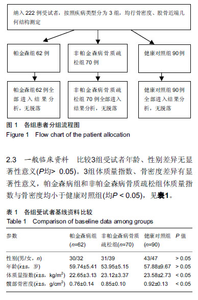
方法:选择抚州市第一人民医院帕金森病患者62例作为帕金森病组,70例非帕金森病骨质疏松者作为非帕金森病骨质疏松组,以90例年龄和性别匹配的健康人群作为健康对照组。所有受试者对试验方案均知情同意,且得到抚州市第一人民医院伦理委员会批准。采用双能X射线骨密度仪及自带的髋部结构分析软件测量骨密度与股骨近端几何结构参数,股骨近端几何结构参数和骨密度的相关性采用Pearson相关分析。
结果与结论:①比较3组受试者股骨颈干角、截面力矩,差异均无显著性意义(P > 0.05);②帕金森病组截面面积、骨皮质厚度小于非帕金森病骨质疏松组及对照组,屈曲应力比大于非帕金森病骨质疏松组及对照组,差异均有显著性意义(P < 0.05);③骨密度与股骨颈、转子间截面面积、骨皮质厚度呈高度正相关(P < 0.01),与屈曲应力比呈高度负相关(P < 0.01),与股骨颈干角、截面力矩无相关性(P均>0.05);④提示帕金森患者骨密度减低,股骨近端结构发生改变,表现为骨截面面积、骨皮质厚度降低,屈曲应力比增高,增加帕金森病患者髋部骨折风险;髋部骨折发生与髋部骨密度及股骨近端几何结构改变有关。


中国组织工程研究杂志出版内容重点:人工关节;骨植入物;脊柱骨折;内固定;数字化骨科;组织工程
ORCID: 0000-0002-5558-8900(王武华)

关键词: 帕金森病, 髋部骨折, 骨密度, 股骨近端几何结构, Pearson相关分析, 股骨颈干角, 截面力矩, 骨皮质厚度

Abstract:

BACKGROUND: Patients with Parkinson’s disease have a high risk of falls and low bone density, so the incidence of fracture increases. It is important to understand the risk factors associated with hip fracture and predict risk.
OBJECTIVE: To investigate the variations of hip bone mineral density and geometric structure of proximal femur in patients with Parkinson’s disease, and to predict the risk for hip fracture.
METHODS: Sixty-two cases of Parkinson’s disease and 70 cases of osteoporotic non-Parkinson’s disease at the First People’s Hospital of Fuzhou as Parkinson’s disease group were collected from the First People’s Hospital of Fuzhou, and 90 healthy controls matched for sex and age were enrolled. All patients assigned the informed consents, and the study was approved by the Ethics Committee of the First People’s Hospital of Fuzhou. The bone mineral density and geometric structure parameters of proximal femur were measured by dual-energy X-ray absorptiometry and hip structural analysis software and their correlations were analyzed by Pearson correlation analysis.
RESULTS AND CONCLUSION: (1) There was no significant difference in the neck shaft angle and cross-sectional moment among groups (P > 0.05). (2) The Parkinson’s disease group had decreased cross sectional area and cortical thickness, and increased upper buckling ratio compared with the non-Parkinson’s disease and healthy control groups (P < 0.05). (3) Bone mineral density showed positive correlation with cross sectional area and cortical thickness, and showed negative correlation with bucking ratio (P < 0.01). Bone mineral density had no correlation with neck shaft angle and cross-sectional moment of inertia both (both P > 0.05). (4) These results indicate that Parkinson’s disease patients have low bone mineral density, decreased cross sectional area and cortical thickness, and high upper buckling ratio, which may increase the risk for hip fracture. Incidence of hip fracture is associated with the bone mineral density of hip and changed geometric structure of proximal femur.

Key words: Parkinson’s disease, hip fracture, bone mineral density, geometric structure of proximal femur, Pearson correlation analysis, femoral neck shaft angle, cross-sectional moment, cortical thickness

中图分类号: 

','1');return false;" target="_blank">
R459.9