| [1] Purcell RL, Cody JP, Gordon W, et al. Outcomes of war related femoral neck fractures. Injury. 2015;46(12):2399-2403.[2] Della RG. Gaps and opportunities in the management of the young femoral neck fracture. Injury. 2015;46(3):515-518.[3] Slobogean GP, Sprague SA, Scott T, et al. Complications following young femoral neck fractures. Injury. 2015;46(3):484-491.[4] Zeng X, Zhan K, Zhang L, et al. The impact of high total cholesterol and high low-density lipoprotein on avascular necrosis of the femoral head in low-energy femoral neck fractures. J Orthop Surg Res. 2017; 12(1):30.[5] Takigawa N, Yasui K, Eshiro H, et al. Clinical results of surgical treatment for femoral neck fractures with the Targon((R)) FN. Injury. 2016;47 Suppl 7:S44-S48.[6] Boss JH, Misselevich I. Osteonecrosis of the femoral head of laboratory animals: the lessons learned from a comparative study of osteonecrosis in man and experimental animals. Vet Pathol. 2003; 40(4):345-354.[7] Kregor PJ. The effect of femoral neck fractures on femoral head blood flow. Orthopedics. 1996;19(12):1031-1036, 1037-1038.[8] Ehlinger M, Moser T, Adam P, et al. Early prediction of femoral head avascular necrosis following neck fracture. Orthop Traumatol Surg Res. 2011;97(1):79-88.[9] Bonnaire F, Schaefer DJ, Kuner E H. Hemarthrosis and hip joint pressure in femoral neck fractures. Clin Orthop Relat Res. 1998;(353): 148-155.[10] Han S, Oh M, Yoon S, et al. Risk stratification for avascular necrosis of the femoral head after internal fixation of femoral neck fractures by Post-Operative Bone SPECT/CT. Nucl Med Mol Imaging. 2017;51(1): 49-57.[11] Ma JX, He WW, Zhao J, et al. Bone microarchitecture and biomechanics of the necrotic femoral head. Sci Rep. 2017;7(1):13345.[12] Ueo T, Tsutsumi S, Yamamuro T, et al. Biomechanical aspects of the development of aseptic necrosis of the femoral head. Arch Orthop Trauma Surg. 1985;104(3):145-149.[13] 懋王,张长青. 股骨头坏死实验动物模型:分类与实验应用[J]. 中国组织工程研究,2014,18(36):5879-5884.[14] Jee WS. D.Sc. (hon) -- one man's association. J Musculoskelet Neuronal Interact. 2006;6(2):113-121.[15] 陈珺,张豪,杨国柱,等. 骨形态计量学目前应用专家共识[J]. 中国骨质疏松杂志,2014,20(9):1031-1038.[16] Min BW, Kim SJ. Avascular necrosis of the femoral head after osteosynthesis of femoral neck fracture. Orthopedics. 2011;34(5):349.[17] Wang T, Sun JY, Zha GC, et al. Analysis of risk factors for femoral head necrosis after internal fixation in femoral neck fractures. Orthopedics. 2014;37(12):e1117-e1123.[18] Ai ZS, Gao YS, Sun Y, et al. Logistic regression analysis of factors associated with avascular necrosis of the femoral head following femoral neck fractures in middle-aged and elderly patients. J Orthop Sci. 2013;18(2):271-276.[19] Zhang YL, Chen S, Ai ZS, et al. Osteonecrosis of the femoral head, nonunion and potential risk factors in Pauwels grade-3 femoral neck fractures: A retrospective cohort study. Medicine (Baltimore). 2016; 95(24):e3706.[20] Wang C, Xu GJ, Han Z, et al. Correlation between residual displacement and osteonecrosis of the femoral head following cannulated screw fixation of femoral neck fractures. Medicine (Baltimore). 2015;94(47):e2139.[21] Lambers FM, Bouman AR, Rimnac CM, et al. Microdamage caused by fatigue loading in human cancellous bone: relationship to reductions in bone biomechanical performance. PLoS One. 2013;8(12):e83662.[22] Ahn AC, Grodzinsky AJ. Relevance of collagen piezoelectricity to "Wolff's Law": a critical review. Med Eng Phys. 2009;31(7):733-741.[23] Gou WL, Lu Q, Wang X, et al. Key pathway to prevent the collapse of femoral head in osteonecrosis. Eur Rev Med Pharmacol Sci. 2015; 19(15):2766-2774.[24] Wang C, Wang Y, Meng H, et al. Microstructure and nanomechanical properties of singletrabecular bone in different Regions of osteonecrosis of the femoral head. J Nanosci Nanotechnol. 2016; 16(3):2264-2269.[25] 孙蕴,贺丽英,马兆坤,等. Ward三角区再研究[J]. 中国骨质疏松杂志, 2016,22(6):706-710.[26] Xiao D, Ye M, Li X, et al. Development of femoral head interior supporting device and 3D finite element analysis of its application in the treatment of femoral head avascular necrosis. Med Sci Monit. 2015;21:1520-1526.[27] Chen Z, Xu Y, Qi Z, et al. The formation and function of the sclerosis rim in the femoral head: A biomechanical point of view. Med Eng Phys. 2015;37(12):1125-1132.[28] Kim JW, Ryu JS, Baek S, et al. The timing of bone SPECT to predict osteonecrosis after internal fixation of femur neck fractures. J Orthop Sci. 2017;22(3):457-462.[29] 张洋,王楠,杨立枫,等. 骨髓间充质干细胞移植联合髓芯减压植骨修复股骨头坏死(英文)[J]. 中国组织工程研究, 2015,19(6):883-890.[30] Yu T, Xie L, Chu F. A sclerotic rim provides mechanical support for the femoral head in osteonecrosis. Orthopedics. 2015;38(5):e374-e379.[31] Escudier JC, Ollivier M, Donnez M, et al. Superimposition of maximal stress and necrosis areas at the top of the femoral head in hip aseptic osteonecrosis. Orthop Traumatol Surg Res. 2018.[32] Yi W, Tian Q, Dai Z, et al. Mechanical behaviour of umbrella-shaped, Ni-Ti memory alloy femoral head support device during implant operation: a finite element analysis study. PLoS One. 2014;9(6):e100765.[33] Ma J, Sun W, Gao F, et al. Porous tantalum implant in treating osteonecrosis of the femoral head: still a viable option? Sci Rep. 2016; 6:28227.[34] Zeng YR, He S, Feng WJ, et al. Vascularised greater trochanter bone graft, combined free iliac flap and impaction bone grafting for osteonecrosis of the femoral head. Int Orthop. 2013;37(3):391-398.[35] Yao C, Yi N, Shen J, et al. Clinical reports of surgical dislocation of the hip with sequestrum clearance and impacting bone graft for grade IIIA-IIIB aseptic necrosis of femoral head (ANFH) patients. Oncotarget. 2017;8(30):50084-50090. |
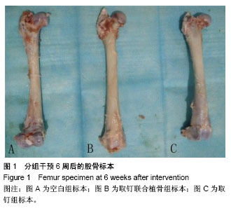
.jpg)

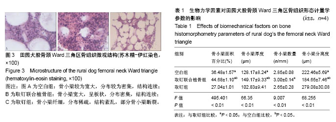
.jpg)