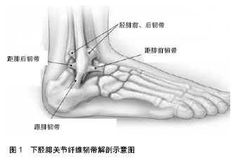
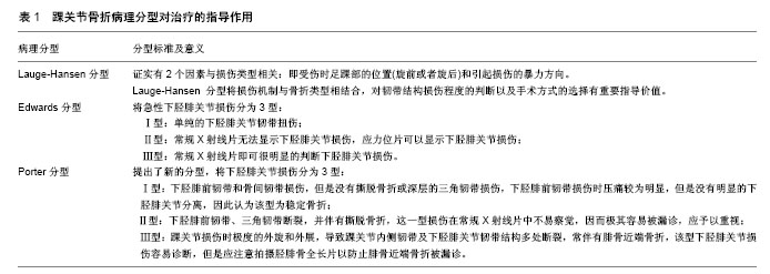
| [1] Bartonicek J. Anatomy of the tibiofibular syndesmosis and its clinical relevance. Surg Radiol Anat.2003; 25:379-386.[2] Beumer A,van Hemert WL,Swierstra BA,et al.A biomechanical evaluation of the tibiofibular and tibiotalar ligaments of the ankle. Foot Ankle Int. 2003;5: 426-429.[3] Porter DA. Ligamentous injuries of the foot and ankle. In: Fitzgerald RH, Kaufer H, Malkani AL, editors. Orthopaedics. St. Louis: Mosby, 2002.[4] Brosky T, Nyland J, Nitz A, et al. The ankle ligaments: consideration of syndesmotic injury and implications for rehabilitation. J Orthop Sports Phys Ther. 1995;21:197-205.[5] Taylor DC, Bassett Iii FH. Syndesmosis ankle sprains: diagnosing the injury and aiding recovery. Phys Sportsmed. 1993;21:39-46.[6] Kjaer MKM, Magnusson P, Engebretsen L, et al. Sports injury: regional considerations. Diagnosis and treatment. Textbook of sports medicine. Oxford: Blackwell Science Ltd, 2003:540-551.[7] Bloemers FW, Bakker FC. Acute ankle syndesmosis injury in athletes. Eur J Trauma. 2006;32:350-356.[8] Evans JM, Schucany WG. Radiological evaluation of a high ankle sprain. Proceedings (Baylor University Medical Center). 2006;19: 402e405.[9] Scranton PE. Isolated syndesmotic injuries: diastasis of the ankle in the athlete. Tech Foot Ankle Surg. 2002;1:88-93.[10] Van Heest TJ, Lafferty PM. Injuries to the ankle syndesmosis. J Bone Joint Surg Am. 2014;96:603-613.[11] Alonso A, Khoury L, Adams R. Clinical tests for ankle syndesmosis injury: reliability and prediction of return to function. J Orthop Sports Phys Ther. 1998;27(4):276-284.[12] Beumer A, Swierstra BA, Mulder PG. Clinical diagnosis of syndesmotic ankle instability: evaluation of stress tests behind the curtains. Acta Orthop Scand. 2002;73(6):667-669.[13] de César PC, Avila EM, de Abreu MR. Comparison of magnetic resonance imaging to physical examination for syndesmotic injury after lateral ankle sprain. Foot Ankle Int. 2011;32(12):1110-1114.[14] Lindenfeld T, Parikh S. Clinical tip: heel-thump test for syndesmotic ankle sprain. Foot Ankle Int. 2005;26(5):406-408.[15] Kiter E, Bozkurt M. The crossed-leg test for examination of ankle syndesmosis injuries. Foot Ankle Int. 2005;26(2):187-188.[16] Lin CF, Gross ML, Weinhold P. Ankle syndesmosis injuries: anatomy, biomechanics, mechanism of injury, and clinical guidelines for diagnosis and intervention. J Orthop Sports Phys Ther. 2006;36(6): 372-384.[17] Press CM, Gupta A, Hutchinson MR. Management of ankle syndesmosis injuries in the athlete. Curr Sports Med Reports. 2009; 8(5):228-233.[18] Takao M, Ochi M, Oae K, et al. Diagnosis of a tear of the tibiofibular syndesmosis. The role of arthroscopy of the ankle. Bone Joint J. 2003;85(3):324-329.[19] Lui TH, Ip KY, Chow HT. Comparison of radiologic and arthroscopic diagnoses of distal tibiofibular syndesmosis disruption in acute ankle fracture.Arthroscopy. 2005;21(11):1370.[20] Lauge-Hansen N. Fractures of the ankle. II. Combined experimentalsurgical and experimental-roentgenologic investigations. Arch Surg. 1950; 60: 957e85.[21] Warner SJ, Garner MR, Hinds RM, et al. Correlation between the Lauge-Hansen Classification and ligament injuries in ankle fractures. J Orthop Trauma. 2015;29(12);574-578.[22] Edwards GS Jr, De Lee JC. Ankle diastasis without fracture. Foot Ankle. 1984;4(6):305-312.[23] Porter DA. Evaluation and treatment of ankle syndesmosis injuries. Instr Course Lect. 2009;58:575-581.[24] Egol KA, Dolan R, Koval KJ. Functional outcome of surgery for fractures of the ankle. A prospective, randomised comparison of management in a cast or a functional brace. J Bone Joint Surg Br. 2000;82:246-249.[25] Amendola A, Williams G, Foster D. Evidence-based approach to treatment of acute traumatic syndesmosis (high ankle) sprains. Sports Med Arthrosc. 2006;14:232-236.[26] Gardner MJ, Graves ML, Higgins TF, et al. Technical considerations in the treatment of syndesmotic injuries associated with ankle fractures. J Am Acad Orthop Surg. 2015;23(8):510-518.[27] Mp VDB, Raven EE. Current concepts review: operative techniques for stabilizing the distal tibiofibular syndesmosis. Foot Ankle Int. 2007; 28(12):1302-1308. [28] Monga P, Kumar A, Simons A, et al. Management of distal tibio-fibular syndesmotic injuries: a snapshot of current practice.. Acta Orthopaedica Belgica. 2008;74(3):365-369.[29] Fallat L, Grimm DJ, Saracco JA. Sprained ankle syndrome: prevalence and analysis of 639 acute injuries.J Foot Ankle Surg. 1998;37:280-285.[30] van den Bekerom MP, Lamme B, Hogervorst M, et al. Which ankle fractures require syndesmotic stabilization? J Foot Ankle Surg. 2007; 46(6):456-463.[31] Magan A, Golano P, Maffulli N, et al. Evaluation and management of injuries of the tibiofibular syndesmosis. Br Med Bull. 2014;111(1): 101-115.[32] Hansen M, Le L, Wertheimer S, et al. Syndesmosis fixation: analysis of shear stress via axial load on 3.5-mm and 4.5-mm quadricortical syndesmotic screws. J Foot Ankle Surg. 2006;45(2):65-69.[33] Markolf KL, Jackson SR, McAllister DR. Syndesmosis fixation using dual 3.5 mm and 4.5 mm screws with tricortical and quadricortical purchase: a biomechanical study. Foot Ankle Int. 2013;34(5):734-739.[34] Stuart K, Panchbhavi VK. The fate of syndesmotic screws. Foot Ankle Int. 2011;32(5):S519-525.[35] Wikeroy AK, Hoiness PR, Andreassen GS, et al. No difference in functional and radiographic results 8.4 years after quadricortical compared with tricortical syndesmosis fixation in ankle fractures. J Orthop Trauma. 2010;24(1):17-23.[36] Jordan TH, Talarico RH, Schuberth JM. The radiographic fate of the syndesmosis after trans-syndesmotic screw removal in displaced ankle fractures. J Foot Ankle Surg. 2011;50(4):407-412.[37] Nousiainen MT, McConnell AJ, Zdero R, et al. The influence of the number of cortices of screw purchase and ankle position in Weber C ankle fracture fixation. J Orthop Trauma. 2008;22(7):473-478.[38] Hoiness P, Stromsoe K. Tricortical versus quadricortical syndesmosis fixation in ankle fractures: a prospective, randomized study comparing two methods of syndesmosis fixation. J Orthop Trauma. 2004;18: 331-337.[39] Moore JA, Shank JR, Morgan SJ, et al. Syndesmosis fixation: a comparison of three and four cortices of screw fixation without hardware removal. Foot Ankle Int. 2006;27(8):567-572.[40] Hahn DM, Colton CL. Malleolar fractures. In: Rüedi TP, Murphy WM (ed). AO Principles of Fracture Management, New York: Thieme, 2000: 559-581.[41] Kukreti S, Faraj A, Miles JN. Does position of syndesmotic screw affect functional and radinterosseous ligamentogical outcome in ankle fractures? Injury. 2005;36:1121-1124.[42] Needleman RL, Skrade DA, Stiehl JB. Effect of the syndesmotic screw on ankle motion. Foot Ankle. 1989;10(1):17-24.[43] Van Heest TJ, Lafferty PM. Injuries to the ankle syndesmosis. J Bone Joint Surg Am. 2014;96:603-613.[44] Hsu YT, Wu CC, Lee WC, et al. Surgical treatment of syndesmotic diastasis: emphasis on effect of syndesmotic screw on ankle function. Int Orthop. 2011;35(3):359-364.[45] Hamid N, Loeffler BJ, Braddy W, et al. Outcome after fixation of ankle fractures with an injury to the syndesmosis: the effect of the syndesmosis screw. J Bone Joint Surg Br. 2009;91:1069-1073.[46] Miller AN, Paul O, Boraiah S, et al. Functional outcomes after syndesmotic screw fixation and removal. J Orthop Trauma. 2010;24(1): 12-16.[47] Schepers T. Complications of syndesmotic screw removal. Foot Ankle Int. 2011.[48] Weening B, Bhandari M. Predictors of functional outcome following transsyndesmotic screw fixation of ankle fractures. J Orthop Trauma. 2005;19(2):102-108.[49] Lloyd J, Elsayed S, Hariharan K, et al. Revisiting the concept of talar shift in ankle fractures. Foot Ankle Int. 2006;27(10):793-796.[50] Sagi HC, Shah AR, Sanders RW. The functional consequence of syndesmotic joint malreduction at a minimum 2-year follow-up. J Orthop Trauma. 2012 l;26(7):439-443.[51] Song D, Lanzi J, Groth A, et al. The effect of syndesmosis screw removal on the reduction of the distal tibiofibular joint [abstract]. Read at the Annual Meeting of the American Academy of Orthopaedic Surgeons; 2012; San Francisco, CA. Paper no. 617.[52] Gardner MJ, Demetrakopoulos D, Briggs SM, et al. Malreduction of the tibiofibular syndesmosis in ankle fractures. Foot Ankle Int. 2006;27(10): 788-792.[53] Miller AN, Carroll EA, Parker RJ, et al. Direct visualization for syndesmotic stabilization of ankle fractures. Foot Ankle Int. 2009;30(5): 419-426.[54] Marmor M, Hansen E, Han HK, et al. Limitations of standard fluoroscopy in detecting rotational malreduction of the syndesmosis in an ankle fracture model. Foot Ankle Int. 2011;32(6):616-622.[55] Mendelsohn ES, Hoshino CM, Harris TG, et al. The effect of obesity on early failure after operative syndesmosis injuries. J Orthop Trauma. 2013;27(4): 201-206.[56] Wukich DK, Kline AJ. The management of ankle fractures in patients with diabetes. J Bone Joint Surg Am. 2008;90(7):1570-1578.[57] Perry MD, Taranow WS, Manoli A 2nd, et al. Salvage of failed neuropathic ankle fractures: use of large-fragment fibular plating and multiple syndesmotic screws. J Surg Orthop Adv. 2005;14(2):85-91.[58] Taylor DC, Englehardt DL, Bassett FH 3rd. Syndesmosis sprains of the ankle. The influence of heterotopic ossification. Am J Sports Med. 1992;20(2):146-150.[59] Droog R,Verhage SM. Incidence and clinical relevance of tibiofibular synostosis in fractures of the ankle which have been treated surgically. Bone Joint J. 2015;97-B:945-949. |
.jpg)

.jpg)