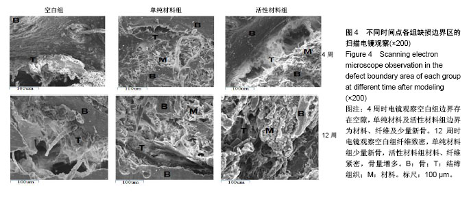
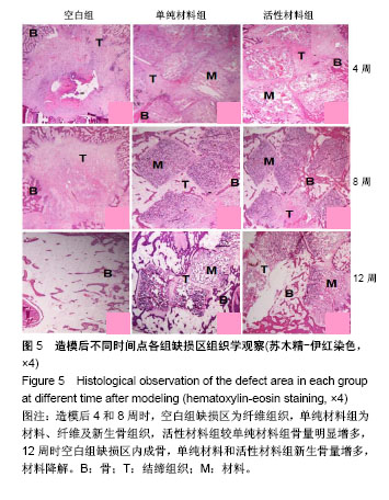
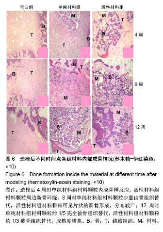
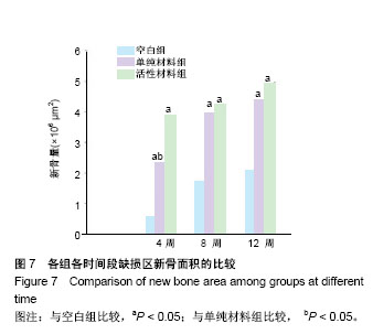
| [1] Amir JA, Abbas A, Simin Z, et al. Mandibular rami implant: a new approach in mandibular reconstruction. J Oral Maxillofac Surg.2017; 75(12):2550-2558.[2] Yang CS, Shen SY, Wu JY, et al. A new modified method for accurate mandibular reconstruction. J Oral Maxillofac Surg. 2018; 11(18): 30-123.[3] 蔡高锐,刘威,何勇,等.纳米生物材料在骨组织工程中的应用[J].国际骨科学杂志,2017, 38(05):303-306.[4] 韩倩倩,薛彬,王涵,等.骨组织工程材料的研究进展[J].组织工程与重建外科杂志,2017, 13(06):343-345.[5] 高伟.同种异体富血小板血浆对兔骨缺损修复作用的实验研究[D]. 河北医科大学. 2017.[6] Harukazu T, Kazunori Y. 141-Growth factors and other new methods for graft-healing enhancement. Elsevier Inc. 2018; 4(3):111-135.[7] Magesh DP, Kumaravelu C, Maheshwari GU. Efficacy of PRP in the reconstruction of mandibular segmental defects using iliac bone grafts. J Maxillofac Oral Surg.2013;12 (2):160-167.[8] Yu T, Pan H, Hu Y, et al. Autologous platelet-rich plasma induces bone formation of tissue-engineered bone with bone marrow mesenchymal stem cells on beta-tricalcium phosphate ceramics. J Orthop Surg Res. 2017; 12(1):178.[9] 綦惠,靳少锋,陈磊,等.富血小板血浆复合再生支架修复兔骨软骨缺损[J].中国矫形外科杂志,2018, 4(08):740-745.[10] 陈骏,张庆福,赵云富,等.富血小板血浆在颌面骨修复中的应用研究进展[J].海军医学杂志,2015, 36(1):93-96.[11] 张喆焱,李晶晶,朱莹,等.富血小板血浆复合羟基磷灰石在即刻种植中骨缺损的应用[J].全科口腔医学电子杂志, 2016, 3(19): 89-90.[12] 朱韵莹,曾国春,武东辉.种植美学区域的富血小板血浆引导骨再生效果评价[J].当代医学,2017, 23(13): 23-24.[13] 钱文慧,徐艳,孙颖,等.富血小板血浆修复牙周骨缺损的临床疗效[J].牙体牙髓牙周病学杂志,2014,24(7):415-418.[14] Dong HL, Keun JR, Jin WK, et al. Bone marrow aspirate concentrate and platelet-rich plasma enhanced bone healing in distraction osteogenesis of the tibia. Clin Orthop Relat Res.2014; 472(12):3789-3797. [15] Xie LN, P, Wang FZ, Cui F, et al. The effect of platelet-rich plasma with mineralized collagen based scaffold on repair of rabbit mandible defect. J Bioact Compat Polym.2010; 25(6): 603-621.[16] 张广德,靳霞,李荣亮,等.BMP-9/EPO双基因共转染ADSCs复合nHAC/PLA支架修复兔下颌骨缺损的实验研究[J].中国口腔颌面外科杂志,2017,15(03):202-208.[17] 王大平,熊建义,朱伟民,等.纳米羟基磷灰石人工骨在骨缺损修复中的临床应用研究[J].中国临床解剖学杂志, 2014, 32(2): 206-209.[18] 滕宇.亲骨性BMP-2活性多肽及其仿生骨修复材料生物活性的实验研究[M].华中科技大学. 2013.[19] 张磊.复合BMP-2来源新型短化纳米骨材料在骨修复中的应用硏究[M].山东大学, 2017.[20] Weimin ZD, Wang JY, Xiong JQ, et al. Study on clinical application of nano-hydroxyapatite bone in bone defect repair. Artif Cells Nanomed Biotechnol. 2015; 43(6):361-365.[21] Salazar VS, Gamer LW, Rosen V. et al. BMP signalling in skeletal development, disease and repair. Nat Rev Endocrinol. 2016; 12(4): 203-221.[22] TMcCarrel TM, Mall NA, Lee AS, et al. Considerations for the use of platelet-rich plasma in orthopedics. Sports Med. 2014; 44(8):1025-1036.[23] 吴珍珍,包崇云,李明政,等.载有人胚胎干细胞的多孔磷酸钙骨水泥与富血小板血浆复合后修复大鼠骨缺损的实验研究[J].中国口腔颌面外科杂志, 2017,15(1):6-10.[24] 王毅,刘杨,杜军,等.富血小板血浆及富血小板血浆/OAM对种植体周围骨缺损修复的实验研究[J].口腔医学,2017, 37(4): 302-306.[25] 李思敏,周晋,颜承伟,等.富血小板血浆缓释载体在骨组织工程中的研究进展[J].口腔医学研究, 2015, 31(6):632-634.637.[26] 付维力,李棋,李箭,等.富血小板血浆在临床骨科中的应用进展[J].中国修复重建外科杂志,2014, 28(10):1311-1316.[27] Mohammad J, Jayachandran M, Rethi M, et al. Effectiveness of platelet rich plasma and bone graft in the treatment of intrabony defects: a clinico-radiographic study. Open Dent J. 2018;12(3):133-154.[28] 潘红娟.富血小板血浆制备技术的优化及其组分的检测[D].吉林大学, 2017.[29] 杨毅,毕鑫,李多玉,等.人工骨材料修复骨缺损:多种复合后的生物学与力学特征[J].中国组织工程研究, 2014,18(16): 2582-2587.[30] Yohei K, Yoshitomo S, Hirofumi N, et al. Leukocyte concentration and composition in platelet-rich plasma influences the growth factor and protease concentrations. J Orthop Sci, 2016; 21(5): 683-689.[31] Vishram S, Rashi S, Gaurav S, et al. Platelet rich plasma-A new revolution in medicine. J Anat Soc India, 2017; 66(4): 127-135. |
.jpg)


.jpg)
.jpg)