中国组织工程研究 ›› 2018, Vol. 22 ›› Issue (10): 1506-1510.doi: 10.3969/j.issn.2095-4344.0707

• 组织工程骨及软骨材料 tissue-engineered bone and cartilage materials • 上一篇    下一篇

钛材料和纳米羟基磷灰石修复大鼠骨缺损:对免疫调控的影响

刘  锋1,梁载明2 
  

  1. 1四川省宜宾市第一人民医院骨一科,四川省宜宾市  644000;2西南医科大学附属医院,四川省泸州市  646000
  • 收稿日期:2018-01-29 出版日期:2018-04-08 发布日期:2018-04-08
  • 作者简介:刘锋,男,1968年生,四川省内江市人,副主任医师,主要从事骨缺损、骨创伤修复研究。
  • 基金资助:
    2016年四川省卫生和计划生育委员会课题项目(16PJ549)

Titanium and nano-hydroxyapatite for bone defect repair in rats: effects on immune regulation

Liu Feng1, Liang Zai-ming2
  

  1. 1Department of Orthopedics, First People’s Hospital of Yibin, Yibin 644000, Sichuan Province, China; 2Affiliated Hospital of Southwest Medical University, Luzhou 646000, Sichuan Province, China
  • Received:2018-01-29 Online:2018-04-08 Published:2018-04-08
  • About author:Liu Feng, Associate chief physician, Department of Orthopedics, First People’s Hospital of Yibin, Yibin 644000, Sichuan Province, China
  • Supported by:
    the Funded Project of Sichuan Provincial Health and Family Planning Committee, No. 16PJ549

摘要:

文章快速阅读:


文题释义:
理想的骨缺损修复材料:应该具备以下特性:具备良好的生物相容性;具有足够的力学性能及良好的生物学适应性;骨传导性能良好且具备诱导性;能为细胞提供足够的增殖空间,具有良好的骨组织界面;可塑性较好。
免疫调节:是指免疫系统中的免疫细胞和免疫分子之间,以及与其他系统如神经内分泌系统之间的相互作用,使得免疫应答以最恰当的形式维持在最适当的水平。免疫调节可比喻为机体的交响乐队,配合好—识别和清除抗原,对自身成分产生免疫耐受,维持内环境的稳定;配合差—病原微生物感染、肿瘤、自身免疫病、免疫缺陷病、超敏反应。
 
背景:研究表明,钛材料具有良好的生物学性能,能调控成骨细胞的黏附、增殖和分化,但有关钛材料在大鼠骨缺损中的修复效果及对机体免疫调控影响的研究较缺乏。
目的:探讨钛材料在大鼠骨质疏松性骨缺损中的修复效果及对机体免疫调控的影响。
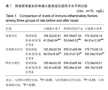
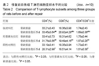
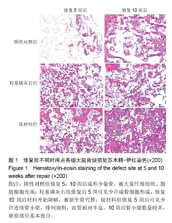
方法:摘除30只Wistar大鼠双侧卵巢,建立骨质疏松模型。3个月后,在30只大鼠股骨远端制备直径为2.5 mm的穿通骨缺损,随机分3组干预,每组10只:阴性对照组不进行任何干预,羟基磷灰石组在骨缺损处填充纳米羟基磷灰石,钛材料组在骨缺损处填充钛材料。干预5 d后,采用酶联免疫吸附实验检测血清白细胞介素1、肿瘤坏死因子α和白细胞介素6水平,采用流式细胞仪检测血清T淋巴细胞亚群水平;干预5,10周后,苏木精-伊红染色观察缺损部位组织形态。
结果与结论:①干预5 d后,钛材料组白细胞介素1、肿瘤坏死因子α和白细胞介素6水平明显低于阴性对照组、羟基磷灰石组(P < 0.05);②干预5 d后,钛材料组CD4+、CD8+、CD4+/CD8+比值均明显高于羟基磷灰石组、阴性对照组(P < 0.05);③苏木精-伊红染色显示,阴性对照组干预5,10周后形成少量骨,被大量纤维组织、脂肪细胞充填;羟基磷灰石组干预5周后可见少许成骨细胞形成,干预10周后材料开始降解,被新生骨代替;钛材料组干预5周后可见少许连续骨小梁,排列规则,血管相对丰富,10周后骨小梁数量较多,缺损部位基本愈合;④结果表明,将钛板材料修复大鼠骨质疏松性骨缺损的效果理想,能调节骨缺损修复中引起的免疫反应。

关键词: 钛材料, 骨质疏松, 骨缺损修复, 纳米羟基磷灰石, 酶联免疫吸附实验, 修复效果, 免疫调控, 生物材料

Abstract:

BACKGROUND: Studies have shown that titanium has good biological properties and regulate osteoblast adhesion, proliferation and differentiation, but there are few reports on the effect of titanium materials on bone defect repair and on immune regulation in rats.
OBJECTIVE: To investigate the effect of titanium materials in the repair of osteoporotic bone defect in rats and its effect on immune regulation.
METHODS: Bilateral ovaries were removed from 30 Wistar rats to establish osteoporosis models. Three months after modeling, a percutaneous bone defect with the diameter of 2.5 mm was prepared on the distal femur of all the 30 rats. These model rats were then randomly divided into three groups with 10 rats in each group: negative control group without any intervention, hydroxyapatite group filled with nano-hydroxyapatite, and titanium material group filled with titanium material. Serum interleukin-1, tumor necrosis factor-α and interleukin-6 levels were measured by enzyme-linked immunosorbent assay after 5 days of intervention, and meanwhile, serum T-lymphocyte subsets were detected by flow cytometry. After 5 and 10 weeks of intervention, hematoxylin-eosin staining was used for morphological observation of the defect site.
RESULTS AND CONCLUSION: (1) After 5 days of intervention, the levels of interleukin-1, tumor necrosis factor-α and interleukin-6 in the titanium material group were significantly lower than those in the negative control and hydroxyapatite groups (P < 0.05). (2) After 5 days of intervention, the levels of CD4+ and CD8+ and the ratios of CD4+/CD8+ in the titanium material group were significantly higher than those in the hydroxyapatite and negative control groups (P < 0.05). (3) Hematoxylin-eosin staining results showed that a small amount of bone tissues filled with a large amount of fibrous tissues and adipocytes formed in the negative control group at 5 and 10 weeks after intervention. A little bone formation was observed in the hydroxyapatite group at 5 weeks after intervention, and the implant began to degrade at 10 weeks and was gradually replaced by new bone tissues. A small amount of trabecular bones enriched with blood vessels arranged orderly in the titanium material group at 5 weeks after intervention; and at 10 weeks, the number of trabecular bone increased and the defect was basically healed. Taken together, the titanium plate can achieve good results in the repair of osteoporotic bone defects in rats, and moreover, it can control the immune response during the bone defect repair.

Key words: Titanium, Osteoporosis, Hydroxyapatites, T-Lymphocyte Subsets, Tissue Engineering

中图分类号: