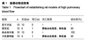
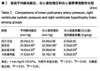
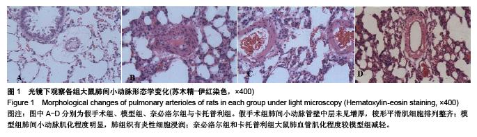
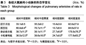
| [1] Neutel JM,Giles TD,Punzi H,et al.Long-term safety of nebivolol and valsartan combination therapy in patients with hypertension: an open-label, single-arm, multicenter study.J Am Soc Hypertens. 2014;8(12):915-920.
[2] Kamp O,Metra M,Bugatti S,et al.Nebivolol: haemodynamic effects and clinical significance of combined beta-blockade and nitric oxide release.Drugs. 2010;70(1):41-56.
[3] Ignarro LJ,Byrns RE,Trinh K,et al.Nebivolol: a selective beta(1)-adrenergic receptor antagonist that relaxes vascular smooth muscle by nitric oxide- and cyclic GMP-dependent mechanisms.Nitric Oxide.2002;7(2): 75-82.
[4] D'Agostino B,Gallellia L,Falciania M,et al.Nebivolol and airway responsiveness in the rabbit.Life Sci.2001; 68(18):2159-2168.
[5] Ocampo C,Ingram P,Ilbawi M,et al.Revisiting the surgical creation of volume load by aorto-caval shunt in rats.Mol Cell Biochem.2003;251(1-2):139-143.
[6] Pinar Yildiz.Molecular mechanisms of pulmonary hypertension. Clinica Chimica Acta 2009, 403: 9-16.
[7] KlingerJR.The nitric oxide/cGMP signaling pathway in pulmonary hypertension.Clin Chest Med. 2007;28(1): 143-167,ix.
[8] Mandegar M,Fung YC,Huang W,et al.Cellular and molecular mechanisms of pulmonary vascular remodeling:role in the development of pulmonary hypertension. Microvasc Res. 2004;68(2):75-103.
[9] Perros F,Ranchoux B,Izikki M,et al.Nebivolol for improving endothelial dysfunction, pulmonary vascular remodeling, and right heart function in pulmonary hypertension.J Am Coll Cardiol. 2015;65(7):668-680.
[10] Vermeersch P,Buys E,Pokreisz P,et al.Soluble guanylate cyclase-alpha1 deficiency selectively inhibits the pulmonary vasodilator response to nitric oxide and increases the pulmonary vascular remodeling response to chronic hypoxia. Circulation. 2007;116(8):936-943.
[11] Napoli C,Paolisso G,Casamassimi A,et al.Effects of nitric oxide on cell proliferation: novel insights. J Am Coll Cardiol. 2013;62(2):89-95.
[12] Sato J,Nair K,Hiddinga J,et al. eNOS gene transfer to vascular smooth muscle cells inhibits cell proliferation via upregulation of p27 and p21 and not apoptosis. Cardiovasc Res.2000;47(4):697-706.
[13] Bakris GL,Basile JN,Giles TD,et al.The role of nitric oxide in improving endothelial function and cardiovascular health: focus on nebivolol.Am J Med. 2010;123(7 Suppl 1):S2-8.
[14] Maffei A,Vecchione C,Aretini A,et al. Characterization of nitric oxide release by nebivolol and its metabolites.BAm J Hypertens.2006;19(6):579-586.
[15] Bayar E,Ilhan G,Furat C,et al.The effect of different β-blockers on vascular graft nitric oxide levels: comparison of nebivolol versus metoprolol.Eur J Vasc Endovasc Surg.2014;47(2): 204-218.
[16] Price A,Raheja P,Wang Z,et al.Differential effects of nebivolol versus metoprolol on functional sympatholysis in hypertensive humans.Hypertension.2013;61:1263-1269.
[17] Valentini M,Revera M,Bilo G,et al.Effects of beta-blockade on exercise performance at high altitude: a randomized, placebo-controlled trial comparing the efficacy of nebivolol versus carvedilol in healthy subjects.Cardiovasc Ther.2012; 30:240-248.
[18] de Nigris F,Rienzo M,Schiano C,et al. Prominent cardioprotective effects of third generation beta blocker nebivolol against anthracycline-induced cardiotoxicity using the model of isolated perfused rat heart.Eur J Cancer.2008; 44(3):334-340.
[19] Wolf SC,Sauter G,Jobst J,et al.Major differences in gene expression in human coronary smooth muscle cells after nebivolol or metoprolol treatment.Int J Cardiol. 2008;125(1): 4-10.
[20] Katragadda S,Arora RR.Role of angiotensin-converting engyme inhibitors in vascular modulation:beyond the hypertensive effects.Am J Ther.2010;17(1):11.
[21] Caglar N,Dincer I.Comparison between nebivolol and ramipril in patients with hypertension and left ventricular hypertrophy: a randomized open blinded end-point (PROBE) trial.Eur Rev Med Pharmacol Sci.2011;15:1359-1368.
[22] Frishman WH.b-Adrenergic blockade in cardiovascular disease.Cardiovasc Pharmacol Ther.2013;18:310-319.
[23] Fares H,Lavie CJ,Ventura HO. Vasodilating versus first -generation b-blockers for cardiovascular protection.Postgrad Med.2012;124:7-15.
[24] Waeber B,Feihl F.Nebivolol and valsartan: useful treatment for hypertension? Lancet.2014;383(6):1864-1866.
[25] Baker JG.The selectivity of β-adrenoceptor agonists at human β1-,β2- and β3-adrenoceptors. Br J Pharmacol. 2010;160: 1048-1061.
[26] Bundkirchen A,Brixius K,Bölck B,et al.Beta 1-adrenoceptor selectivity of nebivolol and bisoprolol. A comparison of [3H]CGP 12.177 and [125I]iodocyanopindolol binding studies.Eur J Pharmacol.2003;460(1):19-26.
[27] Rubin LJ. The Beta-Adrenergic Receptor in Pulmonary Arterial Hypertension:A Novel Therapeutic Target?J Am Coll Cardiol. 2015;65(7):681-683.
[28] Nodari S,Metra M,Dei Cas L.Beta-blocker treatment of patients with diastolic heart failure and arterial hypertension. A prospective, randomized, comparison of the long-term effects of atenolol vs. nebivolol.Eur J Heart Fail.2003;5:621-627.
[29] Conraads VM,Metra M,Kamp O,et al.Effects of the long-term administration of nebivolol on the clinical symptoms, exercise capacity,and left ventricular function of patients with diastolic dysfunction: results of the ELANDD study. Eur J Heart Fail. 2012;14:219-225.
|