中国组织工程研究 ›› 2013, Vol. 17 ›› Issue (18): 3283-3286.doi: 10.3969/j.issn.2095-4344.2013.18.008

• 心肺移植 heart-lung transplantation • 上一篇    下一篇

脑死亡供体肺病理改变及临床移植应用的可行性

农  江1,孙煦勇1,秦  科1,赖彦华1,聂  峰1,董建辉1,文海涛1,李壮江1,丁  祺2,杨建均2,何昌桓2   

  1. 1解放军第三〇三医院移植医学研究院,广西壮族自治区南宁市  530021 
    2解放军第三〇三医院病理科,广西壮族自治区南宁市  530021
  • 收稿日期:2012-09-17 修回日期:2013-03-21 出版日期:2013-04-30 发布日期:2013-04-30
  • 通讯作者: 孙煦勇,解放军第三〇三医院移植医学研究院,广西壮族自治区南宁市 530021 sxywn@sohu.com
  • 作者简介:农江。 675542391@qq.com
  • 基金资助:

    广西科技厅自然科学基金资助(桂科自0991288)。

Pathological changes and clinical application standards of brain-death donor lung

Nong Jiang1, Sun Xu-yong1, Qin Ke1, Lai Yan-hua1, Nie Feng1, Dong Jian-hui1, Wen Hai-tao1, Li Zhuang-jiang1, Ding Qi2, Yang Jian-jun2, He Chang-huan2   

  1. 1 Research Institute of Transplantation Medicine, 303th Hospital of PLA, Nanning  530021, Guangxi Zhuang Autonomous Region, China
    2 Department of Pathology, 303th Hospital of PLA, Nanning  530021, Guangxi Zhuang Autonomous Region, China
  • Received:2012-09-17 Revised:2013-03-21 Online:2013-04-30 Published:2013-04-30
  • Contact: Sun Xu-yong, Research Institute of Transplantation Medicine, 303th Hospital of PLA, Nanning 530021, Guangxi Zhuang Autonomous Region, China sxywn@sohu.com
  • About author:Nong Jiang, Research Institute of Transplantation Medicine, 303th Hospital of PLA, Nanning 530021, Guangxi Zhuang Autonomous Region, China 675542391@qq.com
  • Supported by:

    Natural Science Foundation of Guangxi Science and Technology Bureau, No. Guikezi0991288

摘要:

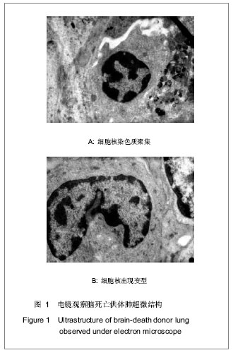
背景:有学者认为在脑死亡过程中很多因素可导致肺组织的改变。
目的:观察脑死亡供体肺病理改变,探讨其临床移植应用的可行性。
方法:对23例脑死亡供体肺进行了病理活检,苏木精-伊红染色、银染及PAS染色观察肺脏组织变化,电镜观察肺脏超微结构变化。
结果与结论:苏木精-伊红、银染及PAS染色光镜下见支气管及肺泡结构尚完整,局部病灶见肺泡内皮细胞水肿、坏死、脱落,肺泡间隔未见明显增宽,但充血易见,血管周围有少量出血,有散在淋巴细胞;电镜下脑死亡的肺泡细胞轻微水肿,部分细胞胞核染色质沿皱缩的核膜下凝聚,有的细胞核出现变型,细胞线粒体肿胀,但未见明显坏死。说明脑死亡供肺可以实施临床移植。

关键词: 器官移植, 器官移植基础实验, 脑死亡, 肺移植, 供体, 省级基金

Abstract:

BACKGROUND: Some scholars believe that many factors can lead to pathological changes of the lung in the process of brain-death
OBJECTIVE: To observe the pathological changes of brain-death donor lung, and to investigate the feasibility of its clinical transplantation.
METHODS: Twenty-three cases of brain-death donor lung received pathological biopsy, and hematoxylin-eosin staining, silver staining and PAS staining were used to observe the pathological changes of lung, and the microstructure of lung tissues was observed under electron microscope.
RESULTS AND CONCLUSION: Under the light microscope, bronchial and alveolar structure was complete, and edema, necrosis and defluxion could be observed in part of epithelial cells by hematoxylin-eosin staining, silver staining and PAS staining; alveolar interval was not widened obviously, but hyperemia was visible; small amount of bleeding and scattered lymphocytes could be seen around the vessels. Under the electron microscope, the slightly edema were observed in the brain-death alveolar cells, the cell nuclear chromatin condensed along the caryotheca, some nucleus appeared abnormity, mitochondria were engorgement, but no cell necrosis could be observed. Brain-death donor lung is suitable for clinical transplantation.

Key words: organ transplantation, basic experiment in organ transplantation, brain death, lung transplantation, donor, provincial grants-supported paper

中图分类号: