中国组织工程研究 ›› 2023, Vol. 27 ›› Issue (18): 2804-2808.doi: 10.12307/2023.298

• 骨与关节生物力学 bone and joint biomechanics • 上一篇    下一篇

肱骨近端骨折中不同腓骨支撑方式重建内侧柱的有限元分析

刘  炎1,葛鸿庆2,陈文治2,梁泽乾3,黎俊烨1,贺杰龙1   

  1. 1广州中医药大学第一附属医院白云医院,广东省广州市   510470;2广东省中医院骨科,广东省广州市   510370;3广州中医药大学第二临床医学院,广东省广州市   510405
  • 收稿日期:2022-04-16 接受日期:2022-05-28 出版日期:2023-06-28 发布日期:2022-09-15
  • 通讯作者: 葛鸿庆,博士,主任医师,广东省中医院骨科,广东省广州市 510370
  • 作者简介:刘炎,男,1993年生,广西壮族自治区北流市人,汉族,2020年广州中医药大学毕业,硕士,医师,主要从事四肢骨折研究。
  • 基金资助:
    广东省中医院横向课题项目(2020KT181),项目负责人:葛鸿庆

Finite element analysis of different fibular support methods to reconstruct the poor medial column of humeral proximal fractures

Liu Yan1, Ge Hongqing2, Chen Wenzhi2, Liang Zeqian3, Li Junye1, He Jielong1   

  1. 1Baiyun Hospital of The First Affiliated Hospital of Guangzhou University of Chinese Medicine, Guangzhou 510470, Guangdong Province, China; 2Department of Orthopaedics, Guangdong Provincial Hospital of Chinese Medicine, Guangzhou 510370, Guangdong Province, China; 3Second Clinical Medical College, Guangzhou University of Chinese Medicine, Guangzhou 510405, Guangdong Province, China
  • Received:2022-04-16 Accepted:2022-05-28 Online:2023-06-28 Published:2022-09-15
  • Contact: Ge Hongqing, MD, Chief physician, Department of Orthopaedics, Guangdong Provincial Hospital of Chinese Medicine, Guangzhou 510370, Guangdong Province, China
  • About author:Liu Yan, Master, Physician, Baiyun Hospital of The First Affiliated Hospital of Guangzhou University of Chinese Medicine, Guangzhou 510470, Guangdong Province, China
  • Supported by:
    Horizontal Project of Guangdong Provincial Hospital of Chinese Medicine, No. 2020KT181 (to GHQ)

摘要:

文题释义:
肱骨近端内侧柱:常称为肱骨内侧距的作用,主要包括肱骨近端后内侧干骺端与内侧铰链(骨膜)对肱骨的支撑作用,临床认为内侧柱粉碎是导致内固定术后不稳定、肱骨头内翻塌陷的常见原因。
有限元分析:是利用数学方法对真实物理系统进行模拟。通过计算机运算模拟将载荷、形态、材料结构和力学性能较为复杂的整体划分为有限个简单的单元,进而充分反映整体内外部应力、应变、位移等力学参数的变化。

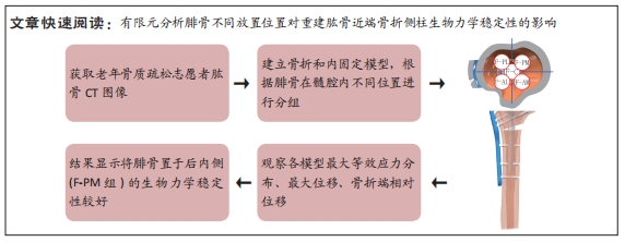
背景:腓骨支撑重建肱骨近端骨折内侧柱已逐渐在临床中广泛应用,而腓骨在髓腔内最佳支撑位点仍存在争议,需要通过生物力学的方法研究其稳定性。
目的:采用有限元分析方法,探讨腓骨在肱骨髓腔中心、前内侧、前外侧、后内侧、后外侧5个位置联合锁定钢板治疗肱骨近端内侧柱缺失型骨折的生物力学稳定性。
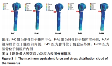
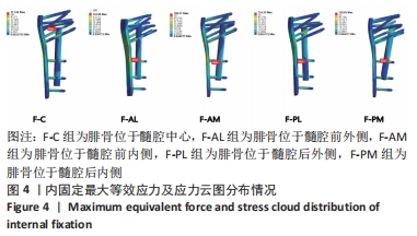
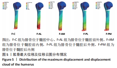
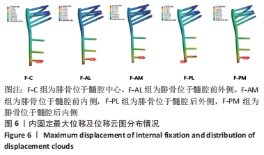
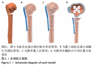
方法:获取1名老年骨质疏松女性患者肱骨近端CT数据,通过Mimics 19.0软件及Geomagic Wrap软件建模,在Soildworks 2017软件中按照肱骨解剖颈下5 mm截骨,建立肱骨近端内侧柱缺失型骨折模型,根据腓骨在髓腔内的位置分为5组:F-C组(腓骨位于髓腔中心)、F-AL组(腓骨位于髓腔前外侧)、F-AM组(腓骨位于髓腔前内侧)、F-PL组(腓骨位于髓腔后外侧)、F-PM组(腓骨位于髓腔后内侧),将数据导入Ansys 2019软件模拟间接暴力状态下不同分组模型的生物力学稳定性。
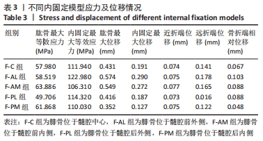
结果与结论:①在600 N轴向载荷下,肱骨应力:F-PL组(49.706 MPa) < F-C组(57.980 MPa) < F-AL组(58.519 MPa) < F-PM组(61.868 MPa) < F-AM组(63.886 MPa),内固定应力:F-AM组(106.310 MPa) < F-PM组(110.030 MPa) < F-C组(111.940 MPa) < F-PL组(114.320 MPa) < F-AL组(122.98 MPa);②在600 N轴向载荷下,肱骨位移:F-PM组(0.352 mm) < F-PL组(0.416 mm) < F-C组(0.431 mm) < F-AM组(0.549 mm) < F-AL组(0.574 mm),内固定位移:F-PM组(0.127 mm) < F-PL组(0.187 mm) < F-C组(0.191 mm) < F-AM组(0.272 mm) < F-AL组(0.290 mm);③骨折端相对位移:F-PM组(0.048 mm)约是F-PL组(0.088 mm)、F-AM组(0.088 mm)的0.54倍,F-C组、F-AL组分别为0.067,0.103 mm;④研究结果表明,F-PM组位移最小,应力较分散,说明将腓骨置于后内侧的生物力学稳定性优于腓骨置于髓腔中心,腓骨置于髓腔外侧的生物力学稳定性最差。

https://orcid.org/0000-0002-5127-7857 (刘炎)

中国组织工程研究杂志出版内容重点:人工关节;骨植入物;脊柱;骨折;内固定;数字化骨科;组织工程

关键词: 肱骨近端骨折, 肱骨近端内固定系统, 植骨, 有限元分析, 生物力学

Abstract: BACKGROUND: The method of fibular allograft to reconstruct the poor medial column of humeral proximal fractures has gradually been widely used in clinic, while the best support site in the intramedullary cavity is still controversial, and it is necessary to study its stability through biomechanical approach.  
OBJECTIVE: To explore the biomechanical stability of the fibula in the center of the medullary cavity, anterolateral, anteromedial, posterolateral, and posteromedial positions using a finite element analysis method.
METHODS:  The proximal humeral CT data of an osteoporosis female patient were obtained, modeled by Mimics 19.0 software and Geomagic Wrap software, and osteotomized in Soildworks 2017 software according to 5 mm below the anatomical neck of the humerus to establish a poor medial column support proximal humeral fracture model. The position of the fibula in the medullary cavity was divided into five groups. The fibula was located in center of the medullary cavity named F-C group; the fibula in anteromedial side of the medullary cavity named F-AL group; the fibula in anteromedial side of the medullary cavity named F-AM group; the fibula in posterolateral side of the medullary cavity named F-PL group; the fibula in posteromedial side of the medullary cavity named F-PM group. The data were imported into Ansys 2019 software to simulate the biomechanical stability of different grouping models under indirect violence.  
RESULTS AND CONCLUSION: (1) Under axial load of 600 N, humeral stress: F-PL group (49.706 MPa)  < F-C group (57.980 MPa) < F-AL group (58.519 MPa) < F-PM group (61.868 MPa) < F -AM group (63.886 MPa); internal fixation stress: F-AM group (106.310 MPa) < F-PM group (110.030 MPa) < F-C group (111.940 MPa) < F-PL group (114.320 MPa) < F-AL group (122.98 MPa). (2) Under 600 N axial loads, humeral deformation: F-PM group (0.352 mm) < F-PL group (0.416 mm) < F-C group (0.431 mm) < F-AM group (0.549 mm) < F-AL group (0.574 mm); internal fixation deformation: F-PM group (0.127 mm) < F-PL group (0.187 mm) < F-C group (0.191 mm) < F-AM group (0.272 mm) < F-AL group (0.290 mm). (3) Relative displacement of the fracture: F-PM group (0.048 mm) was approximately 0.54 times as large as the F-PL group (0.088 mm) and F-AM group (0.088 mm), and the F-C and F-AL groups were 0.067 mm and 0.103 mm. (4) The results of the study showed that the F-PM group had the least displacement and more distributed stresses, indicating that the biomechanical stability of placing the fibula on posteromedial side of the medullary cavity was better than that of placing the fibula in the center of the medullary cavity, and the biomechanical stability of placing the fibula on the lateral side of the medullary cavity was the worst.

Key words: proximal humerus fractures, proximal humeral internal locking system, bone grafting, finite element analysis, biomechanics

中图分类号: