| [1] 戴政,张维军,严翔.慢性根尖周炎根管感染病原菌耐药性分析[J].中华医院感染学杂志,2014,24(12):2920-2922.[2] 纪海,李红,侯本祥.微小小单胞菌与慢性根尖周炎临床症状的相关性研究[J].北京口腔医学,2016,24(1):17-20.[3] 王艳青,胡菊花,李颂,等.亲环素A在人根尖周炎中的表达与临床表现的相关性研究[J].牙体牙髓牙周病学杂志, 2015,25(2): 73-99.[4] 邵丽娜,王丝墨,李晓琳,等.mir146a-5p在慢性根尖周炎病损组织中的表达及临床意义[J].口腔医学研究,2017,33(6),650-653.[5] 徐莉雅,马楠,仇丽鸿.慢性根尖周炎中Wnt5a与骨吸收的关系[J].口腔医学研究,2016,32(2):172-176.[6] 王丽娜,叶丹丹,王娇娇,等.NLRP3/Caspase-1炎性体通路在临床慢性根尖周炎病变组织中的表达[J].大连医科大学学报, 2016, 38(6):538-541.[7] 李燕巍,谢广茹,李玲,等.TLR4/MyD88/NF-κB信号通路对LPS诱导的人鼻咽癌5-8F细胞增殖及凋亡的影响[J].临床耳鼻咽喉头颈外科杂志,2015,29(11):1012-1015.[8] Kogut MH, Iqbal M, He H, et al. Expression and function of Toll-likereceptors in chicken heterophils. Dev CompImmunol. 2005;29(9):791-807. [9] Muzio M, Ni J, Feng P, et al. IRAK (Pelle) family member IRAK-2 and MyD88asproximal mediators of IL-1 signaling. Science.1997;278(5343):1612–1615. [10] Burns K, Martinon F, Esslinger C, et al. MyD88, an adapter protein involved ininterleukin-1signaling.J Chem. 1998;273 (20):12203-12209. [11] Beutler B. Inferences, questions and possibilities in Toll-like receptorSignaling.Nature.2004;430(6996):257-263. [12] Dawen L, Shaomian Y. MyD88 expression in the rat dental forosteoclastogenesisand tooth eruption. Eur J Oral Sci. 2010;118(4):333-341. [13] Lucas K, Maes M. Role of the toll like receptor (TLR) radical cycle in chronic inflammation: possible treatments targeting the TLR4 pathway.MolNeurobiol. 2013;48(1):190-204. [14] 方丽娟,孟民杰.髓样分化因子88研究进展[J].广东药学院学报. 2011,27(2):215-217.[15] 雷升萍,王靓,龙子江,等.黄精多糖通过 TLR4-MyD88-NF-κB通路抑制缺氧/复氧H9c2心肌细胞炎性因子释放[J].中国药理学通报,2017,33(2):255-60.[16] 刘海霞.产黑色素类杆菌与慢性根尖周炎临床症状的相关性[J].牙体牙髓牙周病学杂志,2015,25(9):533-541.[17] Jakovljevic A, Knezevic A, Karalic D, et al. Pro-inflammatorycytokinelevels in human apical periodontitis: Correlation with clinical and histological findings.AustEndod J. 2015;41(2):72-77. [18] 沈苏倩,李娟,黄世光.干细胞因子在人慢性根尖周炎组织中表达的研究[J].口腔医学研究,2017,33(2):179-182.[19] Akira S, Takeda K, Kaisho T. Toll-like receptors: critical proteins linking innate andacquired immunity. Nat Immunol. 2001; 2(8):675–680. [20] JanewayCA. Approaching the asymptote Evolution and revolution inimmunology. Cold Spring Harb Sym. 1989;54: 1-13. [21] Akeuchi O, Kawai T, Sanjo H, et al. TLR6 A novel member of an expandingToll2 like receptor family. Gene.1999; 231(1-2): 59-65,767-769. [22] Medzhitov R, Preston-Hurlburt P, Janeway CA. A human homologue of theDrosop-hila Toll protein signals activation ofadaptive immunity. Nature.1997; 388(6640):394-397. [23] 周毅骏,钦丹萍,杨新艳,等.雷公藤多苷片对溃疡性结肠炎大鼠miR-146a、miR-146b及 TLR4/MyD88依赖信号通路的调控作用研究[J].中草药,2016,47(10):1723-1730.[24] 吴健锋,唐朝霞,陈娟,等.严重脓毒症患者外周血单个核细胞Toll样受体2、4和髓样分化因子88在胸腺肽α1治疗过程中的基因表达变化[J].中华普通外科学文献,2014,8(4 ):270-274.[25] 张超男,黄学宽,骆言,等.电针对急性痛风性关节炎大鼠踝关节滑膜组织TLR/MYD88信号通路的影响[J].四川大学学报, 2014, 45(6):924-927.[26] 孟庆胜,纪健,张丽娟,等.TLR4/MyD88依赖性通路和CaVeolin-1的表达与2型糖尿病神经病变患者炎症状态的相关性[J].临床麻醉学杂志,2015,31(5):449-453.[27] Li YW, Xie GR, Li L.The effect of TLR4/MyD88/NF-κB signaling pathway on proliferation and apoptosis in human nasopharyngeal carcinoma 58F cells induced by LPS.J ClinOtorhinolaryngol Head Neck Surg. 2015;29(11): 1012-1015.[28] 陈秀玲,陈秀慧.髓样分化因88在卵巢癌组织中的表达及其临床意义[J].中国临床药理学杂志,2016,32(24):2250-2256.[29] 李晓杰,董明,牛卫东.大鼠实验性根尖周炎中TLR2表达的研究[J].中国微生态学杂志,2016,7(28):767-769.[30] 胡少婷,黄秦,李升锦.髓样化分子88抑制剂ST2825影响重组耻垢分枝杆菌感染的THP-1细胞炎性分泌功能[J].中国微生态学杂志,2014,10(26):1148-1150.[31] Lin X F, Kong JJ, Wu Q Tl.Effect of TLR4/MyD88 signaling pathway on expression of IL1β and TNF-αin synovia fibroblasts from temporomandibular joint exposed to lipopolysaccharide.Mediators Inflamm. 2015;2015:329405.[32] 王敦方,王彦礼,王怡薇,等.黄芩汤对溃疡性结肠炎大鼠TLR4/MyD88通路调控作用研究[J].药学学报. 2016,51(10): 1558-1563. [33] He W, Yu Q, Zhou Z, et al. CpG oligonucleotides induce an immune response of odontoblasts through the TLR9, MyD88 and NF-kappaB pathways. Biochem Biophys Res Commun. 2010;399(2):274-278.[34] He W, Zhang Y, Zhang J, et al. Cytidine-phosphate-guanosine oligonucleotides ind-uce interleukin-8 production through activation of TLR9, MyD88, NF-κB, and ERK pathways in odontoblast cells. J Endod.2012;38(6):780-5. [35] Zhang J, Zhu QL, Huang P, et al. CpG ODN-induced matrix metalloproteinase-13expression is mediated viaactivation of the ERK and NF-κBsignalling pathways inodontoblast cells. IntEndod J.2013;46(7):666-674. [36] Ogawa T, Hashimoto M, Takeuchi O, et al. Cell activation by Porphyromonasgingivalis lipidAmolecule through Toll-like receptor 4-and myeloid differentia-tion factor88- dependentsignaling pathway. Intimmunol. 2002;14(11): 1325-1332. [37] He W, Qu T, Yu Q, et al. LPS induces IL-8 expression through TLR4, MyD88, NF-kappaB and MAPK pathways in human dental pulp stem cells. Int Endod J.2013;46(2):128-136. [38] Wang L, Jin H, Ye D, et al. Enterococcus faecalisLipoteichoic Acid–induced NLRP3 Inflammasome via the Activation of the Nuclear Factor Kappa B Pathway. J Endod. 2016;42(7): 1093-1100. [39] 梅陵宣,刘正,张濒.实验性鼠根尖周炎组织学动态观察[J].实用口腔医学杂志,2002,18(6):500-503.[40] Wang L, Peng B. Correlation between platelet-derived growth factor B chain andbone resorption in rat periapical lesions. J Endod.2007; 33(6):709–711. |
.jpg) 文题释义:
根尖周炎:指细菌及其分解产物进入到根管系统内从而引起根尖部炎症反应并伴有根尖部牙槽骨破坏的一类疾病。此类炎症是继发于牙髓感染并以持续的抗原刺激引起根尖部牙槽骨组织破坏和宿主反应为主要特征的牙体牙髓疾病。
MyD88:又叫髓样分化因子88,它是Toll/IL-1家族和死亡结构域家族成员。MyD88是参与炎症反应密切相关的TLR2/MyD88信号通路上的重要的衔接蛋白,在信号传递和炎症发生中发挥作用。
摘要
背景:研究发现大鼠牙卵泡中髓样分化因子88有表达,并影响牙齿的萌出和破骨细胞数量。
目的:进一步验证髓样分化因子88在大鼠根尖周炎动物模型中的表达。
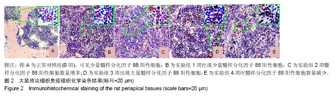
方法:选取6周龄雄性SD大鼠25只,由大连医科大学SPF动物实验中心提供。将大鼠下颌第1磨牙开髓,建立大鼠实验性根尖周炎模型,并将开髓0周作为对照组;开髓1,2,3,4周作为实验组。开髓后于各时间点随机选取5只大鼠,麻醉后取材,用多聚甲醛固定下颌骨,组织脱钙,制作冷冻切片。分别用苏木精-伊红染色、免疫组织化学染色观察根尖组织炎症区域的病理变化,并用RT-PCR检测髓样分化因子88在实验性根尖周炎模型中的表达。
结果与结论:①苏木精-伊红染色:结果显示实验组的根尖区有炎症浸润,并向周围蔓延扩展,表明成功建立了大鼠实验性根尖周炎动物模型;②免疫组织化学染色:结果显示髓样分化因子88在1,2,3,4周4个时间点根尖周炎症区域中均呈阳性表达,髓样分化因子88阳性细胞的表达量在1-3周明显增加,并在第3周时出现高峰;而在4周时,其表达量降低;仅少量髓样分化因子88阳性细胞出现在正常对照组;③RT-PCR结果显示髓样分化因子88 mRNA表达量从1-3周逐渐增高,到第4周下降;④结果说明,在实验性根尖周炎的动物模型中髓样分化因子88有表达,并随时间呈一定的表达趋势,髓样分化因子88可能参与了根尖周炎的发生及根尖牙槽骨的吸收。
中国组织工程研究杂志出版内容重点:组织构建;骨细胞;软骨细胞;细胞培养;成纤维细胞;血管内皮细胞;骨质疏松;组织工程
ORCID: 0000-0001-7925-131X(左美娜)
文题释义:
根尖周炎:指细菌及其分解产物进入到根管系统内从而引起根尖部炎症反应并伴有根尖部牙槽骨破坏的一类疾病。此类炎症是继发于牙髓感染并以持续的抗原刺激引起根尖部牙槽骨组织破坏和宿主反应为主要特征的牙体牙髓疾病。
MyD88:又叫髓样分化因子88,它是Toll/IL-1家族和死亡结构域家族成员。MyD88是参与炎症反应密切相关的TLR2/MyD88信号通路上的重要的衔接蛋白,在信号传递和炎症发生中发挥作用。
摘要
背景:研究发现大鼠牙卵泡中髓样分化因子88有表达,并影响牙齿的萌出和破骨细胞数量。
目的:进一步验证髓样分化因子88在大鼠根尖周炎动物模型中的表达。
方法:选取6周龄雄性SD大鼠25只,由大连医科大学SPF动物实验中心提供。将大鼠下颌第1磨牙开髓,建立大鼠实验性根尖周炎模型,并将开髓0周作为对照组;开髓1,2,3,4周作为实验组。开髓后于各时间点随机选取5只大鼠,麻醉后取材,用多聚甲醛固定下颌骨,组织脱钙,制作冷冻切片。分别用苏木精-伊红染色、免疫组织化学染色观察根尖组织炎症区域的病理变化,并用RT-PCR检测髓样分化因子88在实验性根尖周炎模型中的表达。
结果与结论:①苏木精-伊红染色:结果显示实验组的根尖区有炎症浸润,并向周围蔓延扩展,表明成功建立了大鼠实验性根尖周炎动物模型;②免疫组织化学染色:结果显示髓样分化因子88在1,2,3,4周4个时间点根尖周炎症区域中均呈阳性表达,髓样分化因子88阳性细胞的表达量在1-3周明显增加,并在第3周时出现高峰;而在4周时,其表达量降低;仅少量髓样分化因子88阳性细胞出现在正常对照组;③RT-PCR结果显示髓样分化因子88 mRNA表达量从1-3周逐渐增高,到第4周下降;④结果说明,在实验性根尖周炎的动物模型中髓样分化因子88有表达,并随时间呈一定的表达趋势,髓样分化因子88可能参与了根尖周炎的发生及根尖牙槽骨的吸收。
中国组织工程研究杂志出版内容重点:组织构建;骨细胞;软骨细胞;细胞培养;成纤维细胞;血管内皮细胞;骨质疏松;组织工程
ORCID: 0000-0001-7925-131X(左美娜)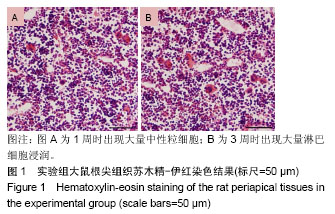


.jpg) 文题释义:
根尖周炎:指细菌及其分解产物进入到根管系统内从而引起根尖部炎症反应并伴有根尖部牙槽骨破坏的一类疾病。此类炎症是继发于牙髓感染并以持续的抗原刺激引起根尖部牙槽骨组织破坏和宿主反应为主要特征的牙体牙髓疾病。
MyD88:又叫髓样分化因子88,它是Toll/IL-1家族和死亡结构域家族成员。MyD88是参与炎症反应密切相关的TLR2/MyD88信号通路上的重要的衔接蛋白,在信号传递和炎症发生中发挥作用。
文题释义:
根尖周炎:指细菌及其分解产物进入到根管系统内从而引起根尖部炎症反应并伴有根尖部牙槽骨破坏的一类疾病。此类炎症是继发于牙髓感染并以持续的抗原刺激引起根尖部牙槽骨组织破坏和宿主反应为主要特征的牙体牙髓疾病。
MyD88:又叫髓样分化因子88,它是Toll/IL-1家族和死亡结构域家族成员。MyD88是参与炎症反应密切相关的TLR2/MyD88信号通路上的重要的衔接蛋白,在信号传递和炎症发生中发挥作用。