| [1]Portan DV,Kroustalli AA,Deligianni DD,et al.On the biocompatibility between TiO2 nanotubes layer and human osteoblasts.J Biomed Mater Res A. 2012;100 (10):2546-2553.
[2]Javed F,Romanos GE.Impact of diabetes mellitus and glycemic control on the osseointegration of dental implants: a systematic literature review.J Periodontol. 2009;80(11):1719-1730.
[3]Cochran DL,Jackson JM,Bernard JP,et al.A 5-year prospective multicenter study of early loaded titanium implants with a sandblasted and acid-etched surface. Int J Oral Maxillofac Implants.2011;26(6):1324-1332.
[4]Faeda RS,Tavares HS,Sartori R,et al.Evaluation of titanium implants with surface modification by laser beam. Biomechanical study in rabbit tibias.Braz Oral Res. 2009;23(2):137-143.
[5]吴明月,周玉琴,李全利,等.钛种植体表面磷酸化与仿生矿化的实验研究[J].中华口腔医学杂志, 2012,47(6): 354-358.
[6]Chellappa M,Anjaneyulu U,Manivasagam G,et al.Preparation and evaluation of the cytotoxic nature of TiO2 nanoparticles by direct contact method.Int J Nanomedicine.2015;10 Suppl 1:31-41.
[7]徐倩,冯青,欧俊,等.层层静电自组装构建载药种植体的研究[J].华西口腔医学杂志, 2014,32(6):537-541.
[8]Das K,Bose S,Bandyopadhyay A.TiO2 nanotubes on Ti: Influence of nanoscale morphology on bone cell-materials interaction.J Biomed Mater Res A. 2009; 90(1):225-237.
[9]Brammer KS,Oh S,Cobb CJ,et al.Improved bone-forming functionality on diameter controlled TiO2 nanotube surface.Acta Biomater. 2009;5(8): 3215-3223.
[10]Song L,Zhao J,Zhang X,et al.Icariin induces osteoblast proliferation, differentiation and mineralization through estrogen receptor-mediated ERK and JNK signal activation.Eur J Pharmacol.2013;714(1-3):15-22.
[11]Xu CQ,Liu BJ,Wu JF,et al.Icariin attenuates LPS-induced acute inflammatory responses: Involvement of PI3K/Akt and NF-kappaB signaling pathway.Eur J Pharmacol.2010;642(1-3):146-153.
[12]Zhao J,Ohba S,Komiyama Y,et al.Icariin: a potential osteoinductive compound for bone tissue engineering.Tissue Eng.2010;16(1):233-243.
[13]Luo Z,Liu M,Sun L,et al.Icariin recovers the osteogenic differentiation and bone formation of bone marrow stromal cells from a rat model of estrogen deficiency-induced osteoporosis.Mol Med Rep.2015; 12(1):382-388.
[14]Gulati K,Ramakrishnan S,Aw MS,et al.Biocompatible polymer coating of titania nanotube arrays for improved drug elution and osteoblast adhesion.Acta Biomater. 2012;8(1):449-456.
[15]Liu X,Li X,Li S,et al.An in vitro study of a titanium surface modified by simvastatin-loaded titania nanotubes-micelles.J Biomed Nanotechnol. 2014; 10(2):194-204.
[16]Zhao J,Ohba S,Shinkai M,et al.Icariin induces osteogenic differentiation in vitro in a BMP- and Runx2-dependent manner.Biochem Biophys Res Commun.2008;369(2):444-448.
[17]Zhang X,Guo Y,Li DX,et al.The effect of loading icariin on biocompatibility and bioactivity of porous β-TCP ceramic.J Mater Sci Mater Med.2011;22(2):371-379.
[18]Geetha M,Singh AK,Asokamani R,et al.Ti based biomaterials, the ultimate choice for orthopaedic implants–A review.Prog Mater Sci.2009;54:397-425.
[19]Beppu K,Kido H,Watazu A,et al.Peri-implant bone density in senile osteoporosis-changes from implant placement to osseointegration.Clin Implant Dent Relat Res.2013;15(2):217-226.
[20]Alghamdi HS,Jansen JA.Bone regeneration associated with nontherapeutic and therapeutic surface coatings for dental implants in osteoporosis. Tissue Eng Part B Rev. 2013;19(3):233-253.
[21]Macak JM,Tsuehiya H,Schmuki P.High-aspect-ratio TiO2 nanotubes by anodization of titanium. Angew Chem Int Ed Engl.2009;44(14):2100-2102.
[22]Oh S,Brammer KS,Li YS,et al.Stem cell fate dictated solely by altered nanotube dimension.Proc Natl Acad Sci USA.2009;106(7):2130-2135.
[23]Das K,Bose S,Bandyopadhyay A,et al.TiO2 nanotubes on Ti:Influence of nanoscale morphology on bone cell-materials interaction.J Biomed Mater Res A. 2009; 90(1):225-237.
[24]Brammer KS,Oh S,Cobb CJ,et al.Improved bone-forming functionality on diameter controlled TiO2 nanotube surface.Acta Biomater.2009;5(8):3215-3223.
[25]于卫强,蒋欣泉,张益琳,等.TiO2纳米管的制备及其对成骨细胞增殖和碱性磷酸酶活性的影响[J].中华口腔医学杂志,2009,44(12):751-755.
[26]Kim JH,Cho KP,Chung YS,et al.The effect of nanotubular titanium surfaces on osteoblast differentiation.J Nanosci Nanotechnol. 2010;10(5): 3581-3585.
[27]Peng Z,Ni J,Zheng K,et al.Dual effects and mechanism of TiO2 nanotube arrays in reducing bacterial colonization and enhancing C3H10T1/2 cell adhesion. Int J Nanomedicine.2013;8:3093-3105.
[28]Yu WQ,Xu L,Zheng K,et al.The effect of Ti anodized nano-foveolae structure on preosteoblast growth and osteogenic gene expression.J Nanosci Nanotechnol. 2014;14(6):4387-4393.
[29]Ma M,Kazemzadeh-Narbat M,Hui Y,et al.Local delivery of antimicrobial peptides using self-organized TiO2 nanotube arrays for peri-implant infections.J Biomed Mater Res A.2012;100(2):278-285.
[30]Zhao L,Wang H,Huo K,et al.Antibacterial nano-structured titania coating incorporated with silver nanoparticles.Biomaterials.2011;32(24):5706-5716.
[31]Ma Y,Zhang Z,Liu Y,et al.Nanotubes Functionalized with BMP2 Knuckle Peptide Improve the Osseointegration of Titanium Implants in Rabbits.J Biomed Nanotechnol. 2015;11(2):236-244.
[32]Li B,Hao J,Min Y,et al.Biological properties of nanostructured Ti incorporated with Ca, P and Ag by electrochemical method.Mater Sci Eng C Mater Biol Appl. 2015;51:80-86.
[33]Moon SH,Lee SJ,Park IS,et al.Bioactivity of Ti-6Al-4V alloy implants treated with ibandronate after the formation of the nanotube TiO2 layer.J Biomed Mater Res B Appl Biomater.2012;100(8):2053-2059.
[34]杨丽,张荣华,朱晓峰,等.淫羊藿苷对大鼠间充质干细胞骨向分化过程中转化生长因子β1、骨形态发生蛋白2 表达的影响[J].中国组织工程研究与临床康复, 2010,14(19): 3518-3522.
[35]Fu SP,Yang L,Hong H,et al.Icariin Promoted Osteogenic Differentiation of SD Rat Bone Marrow Mesenchymal Stem Cells: an Experimental Study.Zhongguo Zhong Xi Yi Jie He Za Zhi. 2015; 35(7):839-846.
[36]Ma HP,Ming LG,Ge BF,et al.Icariin is More Potent Than Genistein in Promoting Osteoblast Differentiation and Mineralization In vitro.J Cell Biochem. 2011;112(3): 916-923.
[37]Ma XN,Zhou J,Ge BF,et al.Icariin induces osteoblast differentiation and mineralization without dexamethasone in vitro.Planta Med. 2013;79(16):1501-1508.
[38]Çal??kan N,Bayram C,Erdal E,et al.Titania nanotubes with adjustable dimensions for drug reservoir sites and enhanced cell adhesion.Mater Sci Eng C Mater Biol Appl. 2014;35:100-105.
[39]Ding X,Zhou L,Wang J,et al.The effects of hierarchical micro/nanosurfaces decorated with TiO2 nanotubes on the bioactivity of titanium implants in vitro and in vivo.Int J Nanomedicine.2015;10:6955-6973.
[40]Jia H,Kerr LL.Sustained ibuprofen release using composite poly(lactic-co-glycolic acid)/titanium dioxide nanotubes from Ti implant surface.J Pharm Sci. 2013; 102(7):2341-2348.
[41]Balasundaram G,Yao C,Webster TJ.TiO2 nanotubes functionalized with regions of bone morphogenetic protein-2 increases osteoblast adhesion.J Biomed Mater Res A.2008; 84(2):447-453.
[42]Popat KC,Eltgroth M,Latempa TJ,et al.Decreased Staphylococcus epidermis adhesion and increased osteoblast functionality on antibiotic-loaded titania nanotubes. Biomaterials.2007;28(32):4880-4888.
[43]Yang W,Yu XC,Chen XY,et al.Pharmacokinetics and tissue distribution profile of icariin propylene glycol-liposome intraperitonea.J Pharm Pharmacol. 2012;64(2):190-198.
[44]Lee YH,Bhattarai G,Park IS,et al.Bone regeneration around N-acetyl cysteine-loaded nanotube titanium dental implant in rat mandible.Biomaterials. 2013; 34(38):10199-21008.
[45]Gulati K,Aw MS,Losic D.Drug-eluting Ti wires with titania nanotube arrays for bone fixation and reduced bone infection.Nanoscale Res Lett.2011;6:571.
[46]Eaninwene G,Yao C,Webster TJ.Enhanced osteoblast adhesion to drug-coated anodized nanotubular titanium surfaces.Int J Nanomedicine. 2008;3(2): 257-264.
[47]Aw MS,Khalid KA,Gulati K,et al.Characterization of drug-release kinetics in trabecular bone from titania nanotube implants.Int J Nanomedicine. 2012;7: 4883-4892.
[48]吴涛,徐俊昌,南开辉,等.淫羊藿苷促进羊骨髓间充质干细胞的增殖和成骨分化[J].中国组织工程研究与临床康复, 2009,13(19):3725-3729.
[49]Wu Y,Xia L,Zhou Y,et al.Icariin induces osteogenic differentiation of bone mesenchymal stem cells in a MAPK-dependent manner.Cell Prolif. 2015;48(3): 375-384.
[50]Zhang J,Li AM,Liu BX,et al.Effect of icarisid II on diabetic rats with erectile dysfunction and its potential mechanism via assessment of AGEs, autophagy, mTOR and the NO-cGMP pathway. Asian J Androl. 2013;15(1):143-148.
[51]Nuschke A,Rodrigues M,Stolz DB,et al.Human mesenchymal stem cells/multipotent stromal cells consume accumulated autophagosomes early in differentiation.Stem Cell Res Ther.2014;5(6):140.
[52]Pantovic A,Krstic A,Janjetovic K,et al.Coordinated time-dependent modulation of AMPK/Akt/mTOR signaling and autophagy controls osteogenic differentiation of human mesenchymal stem cells. Bone.2013;52:7.
[53]Oh S,Daraio C,Chen LH,et al.Significantly accelerated osteoblast cell growth on aligned TiO2 nanotuhes. J Biomed Mater Res A.2006;78(1):97-103.
[54]Minagar S,Wang J,Berndt CC,et al.Cell response of anodized nanotubes on titanium and titanium alloys - A review.J Biomed Mater Res A.2013;101(9):2726-2739.
[55]Shekaran A,García AJ.Extracellular matrix-mimetic adhesive biomaterials for bone repair.J Biomed Mater Res A.2011;96(1):261-272.
[56]曹馨,于卫强,张富强.RGD肽修饰TiO2纳米管对成骨细胞黏附的影响[J].口腔医学研究, 2012,28(6):522-525,530.
[57]Richard M Thomas AE.The biology of fracture healing. Injury.2011;42(6):551-555.
[58]Leighton Y,Carvajal JC,Valenzuela F,et al.Synthesis of nanostructured porous silica coatings on titanium and their cell adhesive and osteogenic differentiation properties.J Biomed Mater Res A.2014;102(1):37-48.
[59]Hsieh TP,Sheu SY,Sun JS,et al.Icariin isolated from Epimedium pubescens regulates osteoblasts anabolism through BMP-2, Smad4, and Cbfa1 expression.Phytomedicine. 2010;17(6):414-423. |
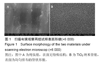
.jpg)
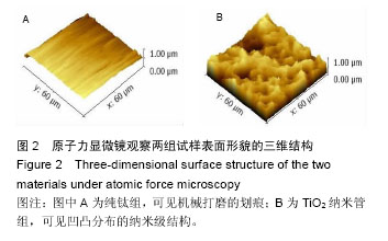
.jpg)
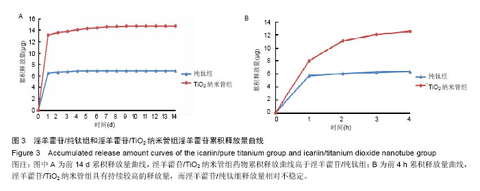
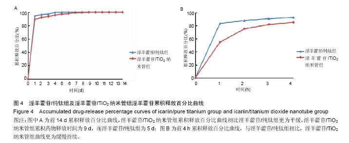
.jpg)