中国组织工程研究 ›› 2016, Vol. 20 ›› Issue (21): 3117-3123.doi: 10.3969/j.issn.2095-4344.2016.21.011

• 细胞外基质材料 extracellular matrix materials • 上一篇    下一篇

脱细胞羊膜与医用膜修复腱鞘缺损防治肌腱粘连的比较

刘国立,于昆仑,白江博,马 韬,杨彦涛,田德虎   

  1. 河北医科大学第三医院,河北省石家庄市  050000
  • 收稿日期:2016-02-24 出版日期:2016-05-20 发布日期:2016-05-20
  • 通讯作者: 田德虎,主任医师,教授,硕士生导师,河北医科大学第三医院手外科,河北省石家庄市 050000
  • 作者简介:刘国立,男,1986年生,河北省唐山市人,汉族,河北医科大学在读硕士,主要从事手外科研究。

 Acellular amniotic membrane versus medical membrane to prevent tendon adhesion in tendon sheath repair

Liu Guo-li, Yu Kun-lun, Bai Jiang-bo, Ma Tao, Yang Yan-tao, Tian De-hu   

  1. (the Third Hospital of Hebei Medical University, Shijiazhuang 050000, Hebei Province, China
  • Received:2016-02-24 Online:2016-05-20 Published:2016-05-20
  • Contact: Tian De-hu, Chief physician, Professor, Master’s supervisor, the Third Hospital of Hebei Medical University, Shijiazhuang 050000, Hebei Province, China
  • About author:Liu Guo-li, Studying for master’degree, the Third Hospital of Hebei Medical University, Shijiazhuang 050000, Hebei Province, China

摘要:

 文章快速阅读:

文题释义:
脱细胞羊膜:
是经处理后的羊膜,去除上皮细胞层,仅有基底膜,因其组织相容性好,抗原性低,无毒副作用,在体内可被降解,在烧伤、眼科、妇产等领域等到了广泛应用。如 Gris等报道了冻干羊膜在眼科疾病治疗方面的作用,患者在随访中无不良反应;董丽霞等成功完成羊膜移植阴道成形37例,术后35例获随访,效果均满意。
理想的腱鞘修补材料:①可被吸收,在体内不残留任何异物。②获取容易,组织相容性好,无局部及全身无不良反应。③具有良好的通透性,在阻断肌腱外源性愈合的同时不影响肌腱愈合质量。④最好含有促进细胞黏附、生长、增殖和分化的因子。

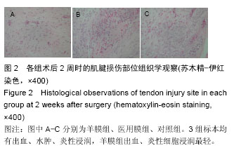
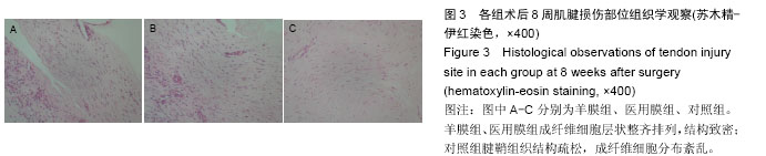
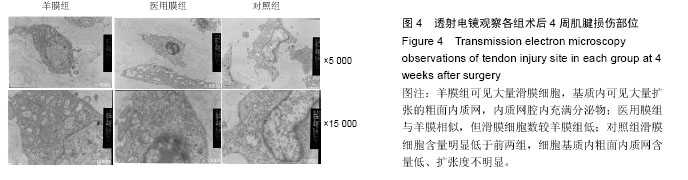
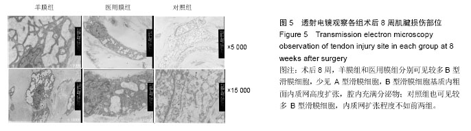
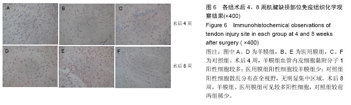
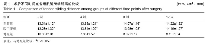
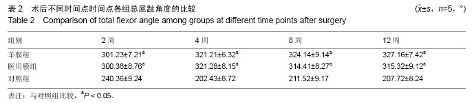
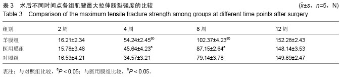
背景:实验证实生物膜可用于腱鞘缺损的重建,抑制肌腱外源性愈合,为肌腱滑动提供良好的基床,减轻肌腱粘连。
目的:比较脱细胞羊膜与医用膜修复腱鞘缺损中防治肌腱粘连的作用。
方法:于66只来亨鸡双足第Ⅲ趾制备肌腱损伤、腱鞘缺损模型,随机分为3 组,每组22只,羊膜组采用脱细胞羊膜修复腱鞘缺损,医用膜组采用可吸收医用膜修复腱鞘缺损,对照组不修复腱鞘。术后2,4,8,12周取各组标本,进行大体、组织学及生物力学检测。
 结果与结论:①大体观察:术后12周,羊膜组、医用膜组腱鞘完整,肌腱愈合良好,无粘连;对照组肌腱粘连严重。②组织学:术后8周,羊膜组假鞘中滑膜细胞数最多,基质内粗面内质网高度扩张,分泌旺盛,医用膜组次之,对照组滑膜细胞数最少,分布杂乱,基质内扩张空泡较前两组弱。术后12
周羊膜组、医用膜组成纤维细胞层状整齐排列,结构致密;对照组腱鞘组织结构疏松,成纤维细胞分布絮乱。③生物力学:羊膜组、医用膜组肌腱滑动距离、总屈趾角度均大于对照组(P < 0.05),前两组间比较差异无显著性意义。术后12周,3组间最大拉伸断裂强度比较差异无显著性意义。④结果表明,脱细胞羊膜与医用膜重建腱鞘缺损均能明显屏障肌腱外源性愈合,防治肌腱粘连。


 ORCID: 0000-0002-9024-1654(田德虎)

关键词: 生物材料, 材料相容性, 脱细胞羊膜, 医用膜, 肌腱粘连, 肌腱愈合

Abstract:

BACKGROUND: Experiments have demonstrated that biological membranes can be used to reconstruct the tendon sheath and inhibit exogenous healing of the tendon. Therefore, these membranes provide a good bed for tendon gliding and reduce tendon adhesion.

OBJECTIVE: To compare the effects of acellular amniotic membrane and medical membrane against tendon adhesion during the repair of tendon sheath defects.
METHODS: Toes III from the bipeds of 66 leghorns were chosen to prepare tendon injury and tendon sheath defect models, which were randomly divided into three groups (n=22 per group). Amnion group were repaired with acellular amniotic membrane, medical membrane group with absorbable membrane, and control group had no treatment on tendon sheath defects. Gross, histological and biomechanical tests of each group were performed at 2, 4, 8, 12 weeks after surgery. 

RESULTS AND CONCLUSION: At 12 weeks after surgery, in the amniotic membrane and medical membrane groups, the tendon sheath formed completely, and the tendon healed well, with no adhesion, but in the control group, there was serious tendon adhesion. At 8 weeks after surgery, the number of synovial cells in the false sheath was highest in the amniotic membrane group sequentially followed by the medical membrane group and control group. In the amniotic membrane group, the rough endoplasmic reticulum expanded highly and secreted exuberantly in the matrix, while in the control group, the synovial cells presented with messy arrangement, and expanded vacuoles in the matrix were weaker than those in the other two groups. At 12 weeks after surgery, fibroblasts were arrayed tidily in layer with dense structure in the medical membrane and amniotic membrane groups; but in the control group, fibroblasts were distributed disorderly with loose structure. Tendon sliding distance and total flexor toe angle in the amniotic membrane and medical film groups were significantly larger than those in the control group (P < 0.05), but there was no significant difference between the medical membrane and amniotic membrane groups. Additionally, the maximum tensile fracture strength had no significant difference among three groups at 12 weeks after surgery. These results indicate that both amniotic membrane and medical membrane can markedly protect the tendon from exogenous healing and adhesion.


Key words: Amnion, Tissue Adhesions, Tissue Engineering

中图分类号: