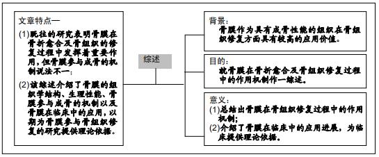
|
[1] SUNG IY, PARK BC, HAH YC, et al. FOXO1 Is Involved in the Effects of Cigarette Smoke Extract on Osteoblastic Differentiation of Cultured Human Periosteum-derived Cells. Int J Med Sci. 2015; 12(11): 881-890.
[2] FILION TM, SKELLY JD, HUANG M, et al. Impaired osteogenesis of T1DM bone marrow-derived stromal cells and periosteum-derived cells and their differential in-vitro responses to growth factor rescue. Stem Cell Res Ther. 2017;8:65.
[3] AUGUSTIN G, ANTABAK A, DAVILA S, et al. The periosteum. Part 1: anatomy, histology and molecular biology. Injury. 2007;38(10):1115- 1130.
[4] 姚作宾,任国良.骨膜血供及其临床意义[J].中国临床解剖学杂志,1991,9(3): 129-131.
[5] 宋守礼,朱盛修. 骨膜的组织学特征和超微结构[J]. 中华骨科杂志, 1996, 16(6):60-62.
[6] BAHNEY CS, HU DP, TAYLOR AJ, et al. Stem cell-derived endochondral cartilage stimulates bone healing by tissue transformation. J Bone Miner Res. 2014;29(5):1269-1282
[7] NETTER FH. Musculoskeletal system: anatomy, physiology, and metabolic disorders. Netter Collection of Medical Illustrations, Saunders, Philadelphia.1988: 171.
[8] 任志勇,李涛,张维彬,等. 兔胫骨干骺端骨膜外截骨对延长区成骨作用的影响[J]. 中国矫形外科杂志, 2015, 23(2):150-155.
[9] ROBERTO-RODRIGUES M, FERNANDES RM, SENOS R, et al. Novel rat model of nonunion fracture with vascular deficit. Injury. 2015; 46(4): 649-654.
[10] TOSCANI D, BOLZONI M, ACCARDI F, et al. The osteoblastic niche in the context of multiple myeloma. Ann N Y Acad Sci. 2015;1335:45-62.
[11] LI C, WEI G, GU Q, et al. Donor Age and Cell Passage Affect Osteogenic Ability of Rat Bone Marrow Mesenchymal Stem Cells. Cell Biochem Biophys. 2015;72(2): 543-549.
[12] 杨丽红,肖波,侯丽霞,等.淫羊藿苷调节细胞增殖作用的研究进展[J]. 医学信息,2019,32(1): 33-36.
[13] SCHANTZ JT, HUTMACHER DW, CHIM H, et al. Induction of ectopic bone formation by using human periosteal cells in combination with a novel scaffold technology. Cell Transplant. 2002; 11(2):125-138.
[14] ZARNETT R, SALTER RB. Periosteal neochondrogenesis for biologically resurfacing joints: its cellular origin. Can J Surg. 1989;32(3):171-174.
[15] 米坤龙,段王平,李兵,等.自体骨膜复合骨髓间充质干细胞移植修复兔关节软骨缺损[J].中国组织工程研究与临床康复,2009,13(46):9066-9070.
[16] MICHAEL D, HOFFMAN, XIE C, et al. The effect of mesenchymal stem cells delivered via hydrogel-based tissue engineered periosteum on bone allograft healing. Biomaterials. 2013;34(35):8887-8898.
[17] 王建钧,陈建庭,唐焕章,等. 珍珠层人工骨修复兔桡骨节段性缺损的放射学评价[J]. 中国临床解剖学杂志,2007,25(6): 673-676.
[18] HOFFMAN MD, BENOIT DSW. Emulating native periosteum cell population and subsequent paracrine factor production to promote tissue engineered periosteum-mediated allograft healing. Biomaterials. 2015; 52:426-440.
[19] MIZUNO D, KAGAMI H, MIZUNO H, et al. Bone regeneration of dental implant dehiscence defects using a cultured periosteum membrane. Clin Oral Implants Res. 2008;19(3):289-294.
[20] 季鸣,刘杰文. 骨外膜剥离对实验性骨折愈合的影响—超微结构研究[J]. 中华骨科杂志,1994,14(8):498-501.
[21] MARUPANTHORN K, TANTRAWATPAN C, KHEOLAMAI P, et al. Bone morphogenetic protein-2 enhances the osteogenic differentiation capacity of mesenchymal stromal cells derived from human bone marrow and umbilical cord. Int J Mol Med. 2017;39(3):654-662.
[22] HISSNAUER TN, NORBERT S, KORNELIA B, et al. Recombinant human bone morphogenetic protein-2 (rhBMP-2) for the treatment of nonunion of the femur in children and adolescents: a retrospective analysis. Bio Med Res Int. 2017; 2017:1-5.
[23] COOK SD, PATRON LP, SALKELD SL, et al. Repair of articular cartilage defects with osteogenic protein-1 (BMP-7) in dogs. J Bone Joint Surg. 2003;85-A Suppl 3:116-123.
[24] TSUJI K, BANDYOPADHYAY A, HARFE BD, et al. BMP2 activity, although dispensable for bone formation, is required for the initiation of fracture healing. Nat Genet. 2006;38(12):1424-1429.
[25] YU L, DAWSON LA, YAN M, et al. BMP9 stimulates joint regeneration at digit amputation wounds in mice. Nat Commun. 2019;10(1): 424.
[26] ZHANG Z, LI L, YANG W, et al. The effects of different doses of IGF-1 on cartilage and subchondral bone during the repair of full-thickness articular cartilage defects in rabbits.Osteoarthritis Cartilage. 2017;25(2):309-320.
[27] SHENG MH, ZHOU XD, BONEWALD LF, et al. Disruption of the insulin-like growth factor-1 gene in osteocytes impairs developmental bone growth in mice.Bone.2013;52(1):133-144.
[28] RITZ MF, GRAUMANN U, GUTIERREZ B, et al. Traumatic spinal cord injury alters angiogenic factors and TGF-beta1 that may affect vascular recovery. Curr Neurovasc Res. 2010;7(4):301-310.
[29] WEN X, LI X, TANG Y, et al. Chondrocyte FGFR3 Regulates Bone Mass by Inhibiting Osteogenesis. J Biol Chem. 2016;291(48): 24912-24921.
[30] TAKEYAMA K, CHATANI M, INOHAYA K, et al. TGF-β2 signaling is essential for osteoblast migration and differentiation during fracture healing in medaka fish. Bone.2016;86: 68-78.
[31] 杨森,王海龙,盛伟斌,等. 骨折合并脊髓损伤患者血清中转化生长因子β1变化:有利于促进骨折愈合[J].中国组织工程研究,2015,19(2):165-169.
[32] HUH JY, CHOI BH, KIM BY, et al. Critical size defect in the canine mandible. Oral Surg Oral Med Oral Pathol Oral Radiol Endod. 2005; 100(3):296-301.
[33] 潘巨利,马金玲,王海涛,等.骨膜下成骨修复小型猪下颌骨节段性缺损的初步研究[J].中华口腔医学研究杂志,2008,2(1):19-25.
[34] SCHMIDT BL, KUNG L, JONES C, et al. Induced osteogenesis by periosteal distraction. J Oral Maxillofac Surg. 2002; 60(10):1170-1175.
[35] SENCIMEN M, AYDINTUG YS, ORTAKOGLU K, et al. Histomorphometrical analysis of new bone obtained by distraction osteogenesis and osteogenesis by periosteal distraction in rabbits. Int J Oral Maxillofac Surg. 2007;36(3):235-242.
[36] RITSILA V, SANTAVIRTA S, ALHOPURO S, et al. Periosteal and perichondral grafting in reconstructive surgery. Clin Orthop Relat Res. 1994;302(8):259-265.
[37] 张大卫,黄东,牟勇,等.游离骨膜瓣联合自体骨治疗胫骨下段粉碎性骨折及骨不连[J].中华显微外科杂志,2015,38(1):88-91.
[38] 严佐发,苏云,马彦明,等.带蒂胫前骨膜瓣联合自体骨治疗距骨缺血坏死的疗效分析[J].实用骨科杂志,2018,24(12):30-32.
[39] SCHÖNMEYR B, CLAVIN N, AVRAHAM T, et al. Synthesis of a tissue-engineered periosteum with acellular dermal matrix and cultured mesenchymal stem cells. Tissue Engineering Part A. 2009;15:1833-1841.
[40] FAN W, CRAWFORD R, XIAO Y. Enhancing in vivo vascularized bone formation by cobalt chloride-treated bone marrow stromal cells in a tissue engineered periosteum model. Biomaterials. 2010;31:3580-3589.
[41] 邢飞,彭静,陈龙,等.组织工程骨膜包被双相陶瓷磷酸钙修复兔尺骨缺损[J].中国组织工程研究,2016,20(47):7006-7013.
[42] NAKAHARA K, HAGA-TSUJIMURA M, IIZUKA T, et al. Periosteum- Induced Bone Formation by Distraction Osteogenesis: Histologic and Microcomputed Tomography Analysis. Int J Oral Maxillofac Implants. 2016; 31(4): 785-792.
[43] FUJIO M, YAMAMOTO A, ANDO Y, et al. Stromal cell-derived factor-1 enhances distraction osteogenesis-mediated skeletal ltissue regeneration through the recruitment of endothelial precursors. Bone. 2011; 49(4): 693-700.
[44] CASAP N, VENEZIA NB, WILENSKY A, et al. VEGF facilitates periosteal distraction-induced osteogenesis in rabbits: a micro-computerized tomography study. Tissue Eng Part A. 2008; 14(2): 247-253.
[45] WINTER LC, WALBOOMERS XF, BUMGARDNER JD, et al. Intermittent versus continuous stretching effects on osteoblast-like cells in vitro. J Biomed Mater Res A. 2003;67(4): 1269-1275.
[46] MOORE DC, LEBLANC CW, MÜLLER R, et al. Physiologic weightbearing increases new vessel formation during distraction osteogenesis: a micro-tomographic imaging study. J Orthop Res. 2003; 21(3): 489-496.
[47] GUO P, ZENG JJ, ZHOU N. Nonvascular trans-port distraction osteogenesis in bone forma-tion and regeneration. Is it an accidental phenomenon? J Craniomaxillofac Surg. 2015;43:21-27.
[48] ZENG JJ, GUO P, ZHOU N, et al. Treatment of large bone defects with a novel biological transport disc in non-vascular transport distraction osteogenesis. Int J Oral Maxillofac Surg. 2016:S0901502715014800.
|