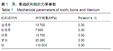
[1] ZHANG Q, WU W, QIAN C, et al. Advanced biomaterials for repairing and reconstruction of mandibular defects. Mater Sci Eng C Mater Biol Appl. 2019. doi: 10.1016/j.msec.2019.109858.
[2] HIDALGO DA. Fibula free flap: a new method of mandible reconstruction. Plastic Reconstruct Surg. 1989;84(1):71-79.
[3] OKAY D, SHETAWI AHA, MOUBAYED SP, et al. Worldwide 10-year systematic review of treatment trends in fibula free flap for mandibular reconstruction. J Oral Maxillofac Surg. 2016;74(12):2526-2531.
[4] 任文豪,高岭,李少明,等.虚拟手术计划及3D打印导板辅助在游离腓骨瓣精准重建下颌骨中的价值分析[J].中华医学杂志,2018, 98(33):2666-2670.
[5] WITJES MJH, SCHEPERS RH, KRAEIMA J. Impact of 3D virtual planning on reconstruction of mandibular and maxillary surgical defects in head and neck oncology. Curr Opin Otolaryngol Head Neck Surg. 2018;26(2):108-114.
[6] AGGARWAL S, SINGH M, MODI P, et al. Comparison of 3D plate and locking plate in treatment of mandibular fracture-a clinical study. Oral Maxillofac Surg. 2017;21(4): 383-390.
[7] KOPP RW, CROZIER DL, GOYAL P, et al. Decade review of mandible fractures and arch bar impact on outcomes of nonsubcondylar fractures. Laryngoscope.2016;126(3): 596-601.
[8] PEDRAZZOLI M, AUTELITANO L, BIGLIOLI F. Prevention of bisphosphonate-related mandibular fractures. Acta Otorhinolaryngol Ital. 2016;36(4):317-320.
[9] JEWER DD, BOYD JB, MANKTELOW RT, et al. Orofacial and mandibular reconstruction with the iliac crest free flap: a review of 60 cases and a new method of classification. Plast Reconstr Surg. 1989;84(3):391-405.
[10] STYRANIVSKA O, KLIUCHKOVSKA N, MYKYYEVYCH N. Comparison of using different bridge prosthetic designs for partial defect restoration through mathematical modeling. Eur J Dent. 2017;11(3):345-351.
[11] OKUDA S, EIRAKU M. Role of molecular turnover in dynamic deformation of a three-dimensional cellular membrane. Biomechan Model Mechanobiol. 2017;16(9):1805-1818.
[12] 杨宇,汤文成.纳米压痕法测量牙周膜不同层面弹性模量(英文)[J].Journal of Southeast University (English Edition), 2017,33(1):33-38.
[13] RUDDY BH, KURUPPUMULLAGE DN, CARNABY G, et al. Computational modelling of cough function and airway penetrant behavior in patients with disorders of laryngeal function. Laryngoscope Invest Otolaryngol. 2017;2(1):23-29.
[14] KUMAR D, SIVARAM G, SHIVAKUMAR B, et al. Comparative evaluation of soft and hard tissue changes following endosseous implant placement using flap and flapless techniques in the posterior edentulous areas of the mandible-a randomized controlled trial. Oral Maxillofac Surg. 2018;22(2):215-223.
[15] MURAKAMI K, YAMAMOTO K, SUGIURA T, et al. Computed tomography-based 3-dimensional finite element analyses of various types of plates placed for a virtually reduced unilateral condylar fracture of the mandible of a patient. J Oral Maxillofac Surg. 2017;75(6):1239.e1-e11.
[16] KANAZAWA M, TANOUE M, MIYAYASU A, et al. The patient general satisfaction of mandibular single-implant overdentures and conventional complete dentures: study protocol for a randomized crossover trial. Medicine. 2018;97(20):e10721.
[17] MAURER P, ECKERT AW, KRIWALSKY MS, et al. Scope and limitations of methods of mandibular reconstruction: a long-term follow-up. Br J Oral Maxillofac Surg. 2010;48(2): 100-104.
[18] DISA JJ, CORDEIRO PG. Mandible reconstruction with microvascular surgery. Semin Surg Oncol. 2000;19(3): 226-234.
[19] SHAYESTEH MOGHADDAM N, JAHADAKBAR A, AMERINATANZI A, et al. Metallic fixation of mandibular segmental defects: graft immobilization and orofacial functional maintenance. Plastic Reconstr Surg. 2016;4(9): e858.
[20] 毛广文,辛俊彤,刘延山,等.双层腓骨移植联合牙种植重建下颌咬合功能[J].现代口腔医学杂志,2016,32(4):221-224.
[21] ZOUMALAN RA, HIRSCH DL, LEVINE JP, et al. Plating in microvascular reconstruction of the mandible: can fixation be too rigid. J Craniofac Surg. 2009;20(5):1451-1454.
[22] STRACKEE SD, KROON FH, BOS KE. Fixation methods in mandibular reconstruction using fibula grafts: acomparative study into the relative strength of three different types of osteosynthesis. Head Neck. 2001;23(1):1-7.
[23] LIU SP, CAI ZG, ZHANG J, et al. Stability and complications of miniplates for mandibular reconstruction with a fibular graft: Outcomes for 544 patients. Br J Oral Maxillofac Surg. 2015; 54(5):496-500.
[24] LI P, SHEN L, LI J, et al. Optimal design of an individual endoprosthesis for the reconstruction of extensive mandibular defects with finite element analysis. J Cranio-Maxillofac Surg. 2014;42(1):73-78.
[25] ALBOGHA MH, MORI Y, TAKAHASHI I. Three-dimensional titanium miniplates for fixation of subcondylar mandibular fractures: comparison of five designs using patient-specific finite element analysis. J Craniomaxillofac Surg. 2018;46(3): 391-397.
[26] STRACKEE SD, KROON FH, BOS KE. Fixation methods in mandibular reconstruction using fibula grafts: a comparative study into the relative strength of three different types of osteosynthesis. Head Neck. 2001;23(1):1-7.
[27] ZHANG ZL, WANG S, SUN CF, et al. Miniplates versus reconstruction plates in vascularized osteocutaneous flap reconstruction of the mandible. J Craniofac Surg. 2019;30(2): e119-e125.
[28] 陈旭兵,柳兆刚,袁建兵,等.三维模拟技术在游离腓骨瓣移植重建下颌骨缺损中的应用[J].上海口腔医学,2015,24(4):460-464.
[29] KOKOSIS G, SCHMITZ R, POWERS DB, et al. Mandibular reconstruction using the free vascularized fibula graft: an overview of different modifications. Arch Plastic Surg. 2016; 43(1):3-9.
[30] 孙伟桐,陈辰,蒋协远,等.合成骨在骨科生物力学研究中的应用[J].国际骨科学杂志,2018,39(2):72-75.
|