中国组织工程研究 ›› 2020, Vol. 24 ›› Issue (4): 566-571.doi: 10.3969/j.issn.2095-4344.2206

• 纳米生物材料 nanobiomaterials • 上一篇    下一篇

磁性靶向纳米粒体外多模态成像及对肝星状细胞的靶向作用

李  璇1,鲁  敏2,李明星1,敖  梦3,4,唐琳梅1,曾  祯1,胡经纬1,黄志强1,宣吉晴1   

  1. 1西南医科大学附属第一医院超声科;2重庆医科大学附属第三医院放射科;3重庆医科大学超声影像学研究所;4重庆医科大学附属第二医院超声科
  • 收稿日期:2019-05-30 修回日期:2019-06-13 接受日期:2019-07-12 出版日期:2020-02-08 发布日期:2020-01-07
  • 通讯作者: 宣吉晴,博士,主治医师,西南医科大学附属第一医院超声医学科,四川省泸州市 646000
  • 作者简介:李璇,女,1994年生,四川省南充市人,西南医科大学附属医院在读硕士,执业医师,主要从事分子影像学研究。
  • 基金资助:
    国家自然科学基金青年基金(81501482);重庆市科委基础学科与前沿技术研究项目(cstc2015jcyjA10045);四川省医学科研青年创新课题(Q17081);四川省泸州市应用基础研究项目(2018-JYJ-44)

In vitro multi-modal imaging of magnetic targeted nanoparticles and their targeting effect on hepatic stellate cells

Li Xuan1, Lu Min2, Li Mingxing1, Ao Meng3, 4, Tang Linmei1, Zeng Zhen1, Hu Jingwei1, Huang Zhiqiang1, Xuan Jiqing1   

  1. 1Department of Ultrasound, The Affiliated Hospital of Southwest Medical University; 2Department of Radiology, The Third Affiliated Hospital of Chongqing Medical University; 3Institute of Ultrasound Imaging, Chongqing Medical University; 4Department of Ultrasound, The Second Affiliated Hospital of Chongqing Medical University
  • Received:2019-05-30 Revised:2019-06-13 Accepted:2019-07-12 Online:2020-02-08 Published:2020-01-07
  • Contact: Xuan Jiqing, MD, Attending physician, Department of Ultrasound, The Affiliated Hospital of Southwest Medical University, Luzhou 646000, Sichuan Province, China
  • About author:Li Xuan, Master candidate, Physician, Department of Ultrasound, The Affiliated Hospital of Southwest Medical University, Luzhou 646000, Sichuan Province, China
  • Supported by:
    the National Natural Science Foundation of China, No. 81501482; Basic Disciplines and Frontier Technology Research Project of Chongqing Science and Technology Commission, No. cstc2015jcyjA10045; Sichuan Medical Research Youth Innovation Project, No. Q17081; Sichuan Luzhou Applied Basic Research Project, No. 2018-JYJ-44

摘要:

文题释义:
多模态成像:随着分子影像学的飞速发展,多模态成像改变了传统单一的成像方式,联合超声、CT、MRI、光声及SPECT等各自独特的成像优势,只使用一种对比剂即可获得多种模态增强显影,同时得到疾病的解剖学、分子学及功能学信息。这对疾病的诊断、受检者的健康及减少医疗资源浪费都有重要意义。
寻靶:通过对微泡外壳进行改建,将特异性配体结合或连接到微泡表面,这些微泡可通过血液循环聚到特定的病变组织上,并长时间停留于靶组织或靶器官,从而达到使病变组织在影像中得到特异性的标记增强或局部靶向治疗作用的目的。

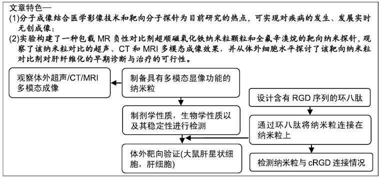
背景:近来年,分子成像结合医学影像技术和靶向分子探针逐渐成为研究的热点,其相互结合能够在分子水平对靶组织进行观察,从而实现对疾病的发生、发展实时无创成像。

目的:制备载磁性颗粒靶向纳米粒探针,探讨其体外超声/CT/MRI成像效果,观察其体外对大鼠肝星状细胞的靶向能力。

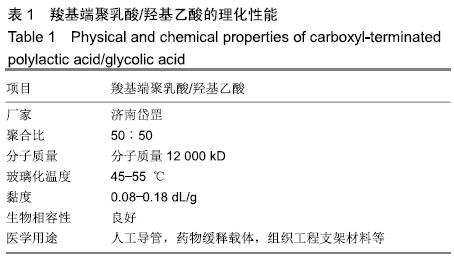
方法:以高分子材料聚乳酸/羟基乙酸作为外壳、含有精氨酸-甘氨酸-天冬氨酸序列环八肽作为配体,通过双步乳化法制备包载磁性颗粒和全氟辛溴烷的载磁性颗粒靶向纳米粒cRGD-PLGA-Fe3O4-PFOB,检测其理化性质。将载磁性颗粒靶向纳米粒以双蒸水稀释为不同质量浓度的混悬液,体外观察其超声、CT、MRI显影效果。通过碳二亚胺法连接精氨酸-甘氨酸-天冬氨酸序列肽与载磁性颗粒靶向纳米粒,验证载磁性颗粒靶向纳米粒的精氨酸-甘氨酸-天冬氨酸序列连接情况和体外靶向能力。细胞毒性实验测定不同质量浓度载磁性颗粒靶向纳米粒对大鼠肝细胞BRL-3A的毒性作用。

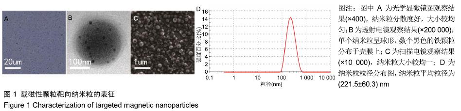
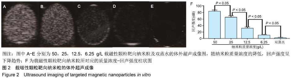
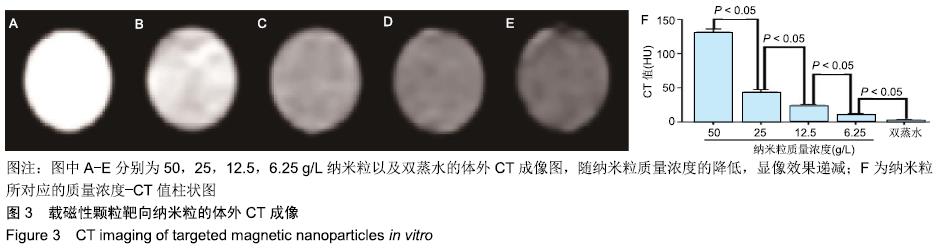
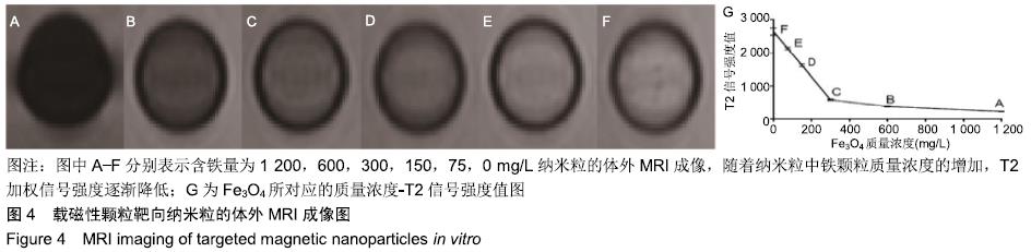
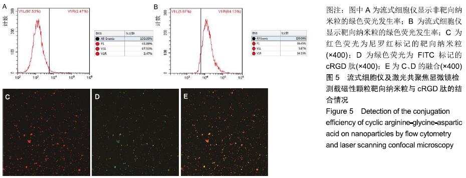
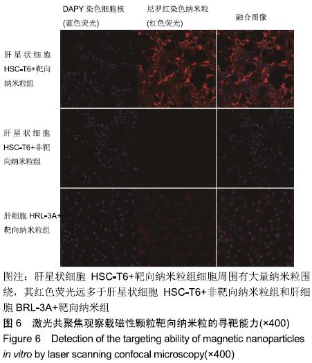
结果与结论:①载磁性颗粒靶向纳米粒分散度好,大小较均匀,单个纳米粒呈球形,数个黑色的铁颗粒分布于壳膜上,平均粒径为(221.5±60.3) nm,Fe3O4包封率为38%;②随着载磁性颗粒靶向纳米粒质量浓度的降低,样品的超声回声强度、CT值均逐渐降低;随着纳米粒中Fe3O4颗粒质量浓度的增加,MRI T2加权信号强度逐渐降低;③流式细胞仪检测显示,含有精氨酸-甘氨酸-天冬氨酸序列环八肽与载磁性颗粒靶向纳米粒的连接率为94.13%;体外靶向实验显示,大部分载磁性颗粒靶向纳米粒聚集于大鼠肝星状细胞细胞HSC-T6周围;④不同质量浓度的载磁性颗粒靶向纳米粒对大鼠肝细胞BRL-3A活力无影响;⑤结果表明,载磁性颗粒靶向纳米粒探针不仅能作为多模态显像剂用于超声、CT、MRI,且在体外实验中对大鼠肝星状细胞有较强的靶向能力,对肝纤维化的早期诊断具有重大应用潜力。

ORCID: 0000-0001-8470-8548(李璇)

中国组织工程研究杂志出版内容重点:生物材料;骨生物材料; 口腔生物材料; 纳米材料; 缓释材料; 材料相容性;组织工程

关键词: 纳米粒子, 靶向, 对比剂, 多模态成像, 肝纤维化, 肝星状细胞, 体外研究, 分子探针

Abstract:

BACKGROUND: In recent years, molecular imaging combined with medical imaging technology and targeted molecular probes have gradually become a research focus. The targeted tissues at the molecular level can be observed using molecular imaging, medical imaging technology, and targeted molecular probes in combination to realize non-invasive imaging of the occurrence and development of the diseases.

OBJECTIVE: To develop the magnetic targeted nanoparticle probes, observe the ultrasound/CT/MRI imaging properties in vitro, and investigate their targeting ability to rat hepatic stellate cells in vitro.

METHODS: Taking poly(lactic-co-glycolic acid) (PLGA) polymer as the shell, cyclic arginine-glycine-aspartic acid (cRGD) octapeptide as the ligand, targeted magnetic nanoparticles with superparamagnetic Fe3O4 embedded in the shell and perfluorooctyl bromide(PFOB) loaded in the core were prepared by double emulsion evaporation method. The physical and chemical properties of the nanoparticles were detected. The ultrasound/CT/MRI multi-modal imaging properties of the nanoparticles at different concentrations diluted with double-distilled water were tested in vitro. Cyclic RGD peptide immobilization on PLGA-Fe3O4-PFOB NPs was completed through the amide condensation reaction. The conjugation efficiency of the cRGD on PLGA-Fe3O4-PFOB NPs and targeting ability of targeted magnetic nanoparticles in vitro were verified. Cytotoxicity experiments were used to measure the toxic effects of nanoparticles at different concentrations on BRL-3A cells in each group.

RESULTS AND CONCLUSION: The targeted magnetic nanoparticles with the average size of (221.5±60.3) nm were uniform in dispersion and size. The prepared individual nanoparticle was spherical with the superparamagnetic Fe3O4 scattered on the shell. The encapsulation rate of Fe3O4 was 38%. In vitro ultrasound imaging and CT imaging signal decreased gradually as the concentrations of the nanoparticle suspension decreased. The T2-weighted signal of MRI decreased gradually with the increase of the concentrations of magnetic particle Fe3O4. Flow cytometry results showed that 94.13% of the cRGD was bound to the nanoparticles. In vitro cell targeting experiments showed that compared to PLGA-Fe3O4-PFOB NPs, cRGD-PLGA-Fe3O4-PFOB NPs exhibited greater cell targeting and affinity efficiency to hepatic stellate cells. Cytotoxicity experiments results showed the nanoparticles had no significant influence on cell viability of the BRL-3A cells. These results suggest that targeted magnetic nanoprobe cannot only be used as a multi-modal imaging contrast agent for ultrasound/CT/MRI, but also exhibits a strong specific affinity to rat hepatic stellate cells in vitro. It has great potential for the early diagnosis of liver fibrosis.

Key words: nanoparticles, target, contrast agent, multi-modal imaging, liver fibrosis, hepatic stellate cells, in vitro, molecular probes

中图分类号: