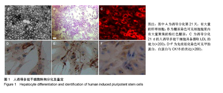
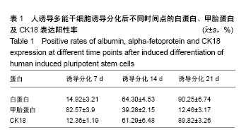
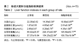
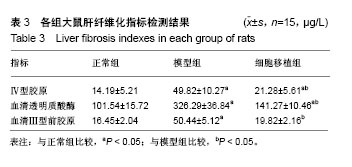
| [1] Zoubek ME, Trautwein C, Strnad P. Reversal of liver fibrosis: From fiction to reality. Best Pract Res Clin Gastroenterol. 2017;31(2):129-141.[2] Pellicoro A, Ramachandran P, Iredale JP. Reversibility of liver fibrosis. Fibrogenesis Tissue Repair. 2012;5(Suppl 1):S26.[3] Pinzani M, Romanelli RG, Magli S. Progression of fibrosis in chronic liver diseases: time to tally the score. J Hepatol. 2001; 34(5):764-767.[4] 唐卫平,胡琦,戴小明,等.脂肪间充质干细胞移植对肝纤维化的治疗作用及其机制[J].中华肝脏病杂志,2018,26(1):28-33.[5] 薛改.脐带间充质干细胞对肝损伤的治疗作用及其机制研究[D].石家庄:河北医科大学,2017.[6] 刘卫,余森源,严和中,等. 骨髓间质干细胞移植对肝纤维化模型大鼠治疗效果[J].临床军医杂志,2017,45(9):903-907.[7] Kim JH, Jang YJ, An SY, et al. Enhanced Metabolizing Activity of Human ES Cell-Derived Hepatocytes Using a 3D Culture System with Repeated Exposures to Xenobiotics. Toxicol Sci. 2015;147(1):190-206.[8] Imai K,Tanoue A,Nakamura K,et al.An attempt to in vitro embryotoxicity test using mouse ES cells and human hepatocytes. Aatex.2013;18(1):33-38.[9] 杜盼,余江南,徐希明.人源多能干细胞体外定向诱导分化为肝细胞[J]. 江苏大学学报(医学版),2018,28(1):39-46.[10] 史德宝,吕礼应. 血清白蛋白检测方法对糖化白蛋白的影响[J].安徽医科大学学报,2015,50(12):1805-1808.[11] 王杨,谷冬梅,陈京山,等. 三种常规方法检测血清胆红素结果的可比性和偏倚评估[J]. 中国实验诊断学, 2017,21(10): 1744-1747.[12] 饶雅琪,张丽,曾金祥,等. 禾叶风毛菊醇提物对四氯化碳致小鼠肝损伤的保护作用[J]. 中国临床药理学杂志, 2017,33(20): 2058-2061.[13] 易梦秋,余旻,王鹏.多器官功能障碍综合征大鼠肺组织MMP-9、TIMP-1和Ⅳ型胶原的表达[J]. 中国病理生理杂志, 2018,34(1): 123-129.[14] Takahashi K, Yamanaka S. Induction of pluripotent stem cells from mouse embryonic and adult fibroblast cultures by defined factors. Cell. 2006;126(4):663-676.[15] Takahashi K, Tanabe K, Ohnuki M, et al. Induction of pluripotent stem cells from adult human fibroblasts by defined factors. Cell. 2007;131(5):861-872.[16] Song Z, Cai J, Liu Y, et al. Efficient generation of hepatocyte-like cells from human induced pluripotent stem cells. Cell Res. 2009;19(11):1233-1242.[17] Twaroski K, Mallanna SK, Jing R, et al. FGF2 mediates hepatic progenitor cell formation during human pluripotent stem cell differentiation by inducing the WNT antagonist NKD1. Genes Dev. 2015;29(23):2463-2474.[18] Takagi C, Yagi H, Hieda M, et al. Mesenchymal Stem Cells Contribute to Hepatic Maturation of Human Induced Pluripotent Stem Cells. Eur Surg Res. 2017;58(1-2):27-39.[19] Mallanna SK, Cayo MA, Twaroski K, et al. Mapping the Cell-Surface N-Glycoproteome of Human Hepatocytes Reveals Markers for Selecting a Homogeneous Population of iPSC-Derived Hepatocytes. Stem Cell Reports. 2016;7(3): 543-556.[20] 刘国菊,李丛丛,李睿坤,等.转化生长因子β1在纤维化疾病发病中作用的研究进展[J].山东医药, 2018,58(30):106-109.[21] 李梓豪,尹大龙,崔逸峰,等.肝硬化肝癌肝部分切除术后影响肝再生的抗纤维化因子研究[J].国际外科学杂志, 2018,45(2): 134-137.[22] 段佳悦,史清峰,王明光,等. 血管内皮生长因子、碱性成纤维细胞生长因子和内皮抑素在肝硬化中的表达及意义[J].临床荟萃, 2018,33(5):427-430.[23] Sulzbacher S, Schroeder IS, Truong TT, et al. Activin A-induced differentiation of embryonic stem cells into endoderm and pancreatic progenitors-the influence of differentiation factors and culture conditions. Stem Cell Rev. 2009;5(2):159-173.[24] 李毅,李欣,孙涛. CDX2基因修饰的脐带间充质干细胞向杯状细胞分化的可行性研究[J].生物技术通讯,2014,25(4):515-519.[25] Kang SJ, Lee HM, Park YI, et al. Chemically induced hepatotoxicity in human stem cell-induced hepatocytes compared with primary hepatocytes and HepG2. Cell Biol Toxicol. 2016;32(5):403-417.[26] 张文成.细胞重编程与诱导内胚层多能干细胞的获取与鉴定[D].北京:中国人民解放军军事医学科学院,解放军军事医学科学院, 2012.[27] Boulter L, Govaere O, Bird TG, et al. Macrophage-derived Wnt opposes Notch signaling to specify hepatic progenitor cell fate in chronic liver disease. Nat Med. 2012;18(4): 572-579.[28] Gieseck RL 3rd, Hannan NR, Bort R, et al. Maturation of induced pluripotent stem cell derived hepatocytes by 3D-culture. PLoS One. 2014;9(1):e86372.[29] Chen Y, Li Y, Wang X, et al. Amelioration of Hyperbilirubinemia in Gunn Rats after Transplantation of Human Induced Pluripotent Stem Cell-Derived Hepatocytes. Stem Cell Reports. 2015;5(1):22-30.[30] Roy-Chowdhury N, Wang X, Guha C, et al. Hepatocyte-like cells derived from induced pluripotent stem cells. Hepatol Int. 2017;11(1):54-69.[31] Sakurai F, Mitani S, Yamamoto T, et al. Human induced-pluripotent stem cell-derived hepatocyte-like cells as an in vitro model of human hepatitis B virus infection. Sci Rep. 2017;7:45698.[32] Huang CS, Lin HC, Lu KH, et al. Generation of high quality of hepatocyte-like cells from induced pluripotent stem cells with Parp1 but lacking c-Myc. J Chin Med Assoc. 2018;81(10): 871-877.[33] Cao YY, Zeng X. Research advances in hepatocyte-like cells from human induced pluripotent stem cells and their application. Zhonghua Gan Zang Bing Za Zhi. 2018;26(1):69-72.[34] Starokozhko V, Hemmingsen M, Larsen L, et al. Differentiation of human-induced pluripotent stem cell under flow conditions to mature hepatocytes for liver tissue engineering. J Tissue Eng Regen Med. 2018;12(5): 1273-1284.[35] Takayama K, Akita N, Mimura N, et al. Generation of safe and therapeutically effective human induced pluripotent stem cell-derived hepatocyte-like cells for regenerative medicine. Hepatol Commun. 2017;1(10):1058-1069.[36] Lorvellec M, Scottoni F, Crowley C, et al. Mouse decellularised liver scaffold improves human embryonic and induced pluripotent stem cells differentiation into hepatocyte-like cells. PLoS One. 2017;12(12):e0189586.[37] Kotaka M, Toyoda T, Yasuda K, et al. Adrenergic receptor agonists induce the differentiation of pluripotent stem cell-derived hepatoblasts into hepatocyte-like cells. Sci Rep. 2017;7(1):16734.[38] Asgari S, Moslem M, Bagheri-Lankarani K, et al. Differentiation and transplantation of human induced pluripotent stem cell-derived hepatocyte-like cells. Stem Cell Rev. 2013;9(4):493-504. |
.jpg)
.jpg)