中国组织工程研究 ›› 2017, Vol. 21 ›› Issue (20): 3117-3122.doi: 10.3969/j.issn.2095-4344.2017.20.001

• 骨组织构建 bone tissue construction •    下一篇

黄精多糖对RANKL诱导骨髓巨噬细胞向破骨细胞分化及体内骨吸收功能的影响

何基琛1,宗少晖1,曾高峰2,杜  力1,彭小明1,施雄智1,吴云乐1   

  1. 1广西医科大学第一附属医院脊柱骨病外科,广西壮族自治区南宁市530021;2广西医科大学公共卫生学院,广西壮族自治区南宁市  530021
  • 收稿日期:2017-02-14 出版日期:2017-07-18 发布日期:2017-07-28
  • 通讯作者: 曾高峰,教授,博士生导师,广西医科大学公共卫生学院,广西壮族自治区南宁市 530021
  • 作者简介:何基琛,男,1990年生,广西壮族自治区南宁市人,壮族,广西医科大学在读硕士,主要从事脊柱外科学方面的研究。
  • 基金资助:

    国家自然科学基金资助项目(81360279)

Polygonatum sibiricum polysaccharide attenuates bone marrow-derived macrophages to differentiate into osteoclasts and protects against lipopolysaccharide-induced osteolysis in vivo

He Ji-chen1, Zong Shao-hui1, Zeng Gao-feng2, Du Li1, Peng Xiao-ming1, Shi Xiong-zhi1, Wu Yun-le1   

  1. 1Department of Spine Osteopathia, the First Affiliated Hospital of Guangxi Medical University, Nanning 530021, Guangxi Zhuang Autonomous Region, China; 2College of Public Health of Guangxi Medical University, Nanning 530021, Guangxi Zhuang Autonomous Region, China
  • Received:2017-02-14 Online:2017-07-18 Published:2017-07-28
  • Contact: Zeng Gao-feng, Professor, Doctoral supervisor, College of Public Health of Guangxi Medical University, Nanning 530021, Guangxi Zhuang Autonomous Region, China
  • About author:He Ji-chen, Studying for master’s degree, Department of Spine Osteopathia, the First Affiliated Hospital of Guangxi Medical University, Nanning 530021, Guangxi Zhuang Autonomous Region, China
  • Supported by:

    the National Natural Science Foundation of China, No. 81360279

摘要:

文章快速阅读:

文题释义:
破骨细胞分化模型:目前应用较为广泛的破骨细胞体外分化模型主要包括RANKL诱导的RAW264.7细胞向破骨细胞分化模型、RANKL和巨噬细胞集落刺激因子共同诱导的小鼠骨髓巨噬细胞向破骨细胞分化模型以及RANKL和巨噬细胞集落刺激因子共同诱导的人外周血单个核细胞向破骨细胞分化模型。骨髓巨噬细胞属于原代破骨细胞前体细胞,在RANKL和巨噬细胞集落刺激因子共同作用下可分化为具有骨吸收功能的破骨样多核细胞。RANKL和巨噬细胞集落刺激因子共同诱导的骨髓巨噬细胞向破骨细胞分化模型属于原代细胞培养模型,对于破骨细胞分化的作用观察及机制研究更具优势。
骨溶解模型:在研究体内骨溶解的防治过程中,动物模型起到了十分重要的作用,建立骨溶解动物模型的方法多种多样,分别为:①假体周围骨溶解模型:包括髋关节骨溶解模型和膝关节骨溶解模型;②气囊(Air-pouch)模型;③颅骨骨溶解模型。大、小鼠颅骨骨溶解模型拥有简单、经济、重复性好的优点,可在较短的时间内观察到药物的防治作用,且个体差异小,具有说服力,可比较广泛的应用到骨溶解防治的研究中。

摘要
背景:
骨髓巨噬细胞具有向破骨细胞分化的潜能。黄精多糖可能抑制骨髓巨噬细胞向破骨细胞分化,有望成为治疗骨质疏松的新药物。
目的:探讨黄精多糖对核因子κ B受体活化因子配体(RANKL)诱导的小鼠骨髓巨噬细胞向破骨细胞分化及体内骨吸收的影响。
方法:培养小鼠骨髓巨噬细胞,在巨噬细胞集落刺激因子和不同质量浓度黄精多糖(5,10,20,40,80,160,320,640,1 280,2 560 mg/L)干预下,CCK-8法检测黄精多糖对小鼠骨髓巨噬细胞增殖的影响以确定黄精多糖质量浓度范围;在巨噬细胞集落刺激因子和RANKL诱导下给予5,10,20,40,80,160,320,640 mg/L黄精多糖进行干预,不加黄精多糖作为对照组,显微镜下观察细胞形态变化。抗酒石酸酸性磷酸酶染色鉴定破骨细胞数目;实时荧光定量PCR检测破骨相关基因抗酒石酸酸性磷酸酶、金属基质蛋白酶9、组织蛋白酶K、活化T细胞核因子c1 mRNA表达。建立脂多糖诱导小鼠颅骨骨溶解模型并给予黄精多糖干预,采用Micro-CT扫描及相关软件定量分析骨体积/组织体积百分比、骨小梁数目、骨小梁间距、骨小梁厚度,并制备组织切片抗酒石酸酸性磷酸酶染色鉴定破骨细胞数目及定量分析骨吸收面积。
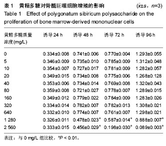
结果与结论:①与对照组相比,质量浓度在640 mg/L以下的黄精多糖对小鼠骨髓巨噬细胞增殖无明显影响(P > 0.05);②不同质量浓度的(40-640 mg/L)黄精多糖可不同程度的减少破骨细胞数目(P < 0.01),显著降低了破骨细胞的分化成熟,同时明显降低抗酒石酸酸性磷酸酶、金属基质蛋白酶9、组织蛋白酶K、活化T细胞核因子c1基因的表达(P < 0.05);③与脂多糖组相比,黄精多糖能有效缓解脂多糖诱导的颅骨骨溶解,骨体积/组织体积百分比、骨小梁数目、骨小梁间距均减小(P < 0.05),破骨细胞数目及骨吸收面明显减少(P < 0.01);④说明黄精多糖能够抑制小鼠骨髓巨噬细胞向破骨细胞分化成熟及缓解脂多糖诱导的颅骨骨溶解。

中国组织工程研究杂志出版内容重点:组织构建;骨细胞;软骨细胞;细胞培养;成纤维细胞;血管内皮细胞;骨质疏松组织工程
ORCID: 0000-0002-0595-0043(曾高峰)

关键词: 组织构建, 骨组织工程, 黄精多糖, 破骨细胞, 颅骨骨溶解模型, 骨质疏松, 国家自然科学基金

Abstract:

BACKGROUND: Bone marrow-derived mononuclear cells (BM-MNCs) hold the potential of differentiating into osteoclasts. Polygonatum sibiricum polysaccharide (PSP) may inhibit the differentiation of BM-MNCs into osteoclasts and it is expected to become a new drug for the treatment of osteoporosis.
OBJECTIVE: To investigate the effect of PSP on the differentiation of mouse BM-MNCs into osteoclasts induced by receptor activator of nuclear factor kappa-B ligand (RANKL) and bone resorption in vivo.
METHODS: Mouse bone marrow-derived macrophages cultured in vitro, the effect of macrophage colony stimulating factor and PSP (5, 10, 20, 40, 80,160, 320, 640, 1 280, 2 560 mg/L) on the proliferation of mouse BM-MNCs was detected by cell counting kit-8 assay to determine the PSP concentration range; the mouse BMMs were cultured and induced in DMEM medium containing macrophage colony stimulating factor, RANKL and 5, 10, 20, 40, 80,160, 320, 640 mg/L PSP, respectively; those cultured without PSP served as control group. The morphological changes of cells were observed under an inverted microscope.; the number of osteoclasts was detected by tartrate-resistant acid phosphatase staining; the mRNA expression levels of osteoclast-related genes including tartrate-resistant acid phosphatase, matrix metalloproteinase-9, cathepsin K, and nuclear factor of activated T cells c1 were evaluated by quantitative real-time PCR. A mouse model of calvarial osteolysis induced by lipopolysaccharide was established to receive PSP intervention, and then micro CT scanning, three-dimensional reconstruction and relevants software were used for quantitative analysis of bone volume/volume percentage, trabecular number, trabecular bone spacing and thickness. The number of osteoclasts was identified by tartrate-resistant acid phosphatase staining and quantitative analysis of bone resorption area was conducted.
RESULTS AND CONCLUSION: Compared with the control group, the concentration of PSP below 640 mg/L showed no significant effect on the proliferation of BMMs (P > 0.05). Different concentrations of PSP (40-640 mg/L) significantly reduced the number of osteoclasts, osteoclast differentiation and maturation, and the mRNA expression levels of tartrate-resistant acid phosphatase, matrix metalloproteinase-9, cathepsin K, and nuclear factor of activated T cells c1 TRAP, MMP-9, CtsK and NFATc1 (P < 0.05). Compared with lipopolysaccharide, PSP could effectively alleviate the lipopolysaccharide-induced calvarial osteolysis, and the bone volume/volume percentage, trabecular number, and trabecular bone spacing were significantly decreased (P < 0.05); additionally, the number of osteoclasts and the area of bone resorption were decreased significantly (P < 0.01). To conclude, PSP can inhibit the differentiation and maturation of mouse BMMs to osteoclasts and alleviate lipopolysaccharide-induced calvarial osteolysis.

中国组织工程研究杂志出版内容重点:组织构建;骨细胞;软骨细胞;细胞培养;成纤维细胞;血管内皮细胞;骨质疏松组织工程

Key words: Polygonatum, Cell Differentiation, Osteoclasts, Osteoporosis, Tissue Engineering

中图分类号: