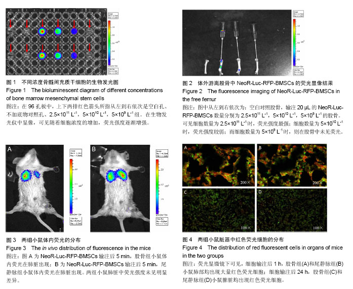
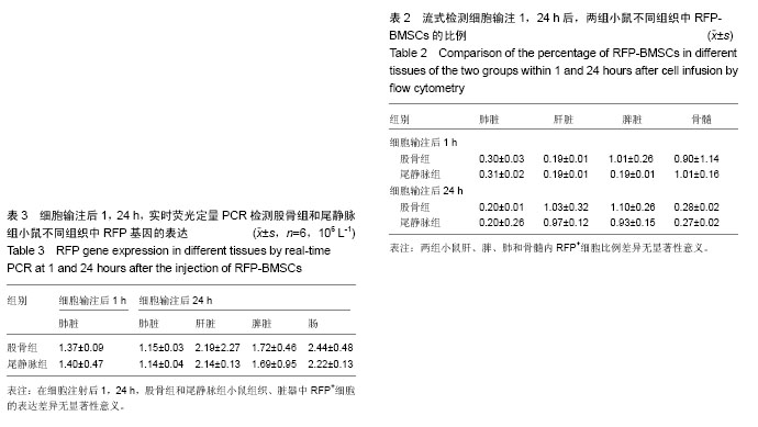
| [1] Auletta JJ, Deans RJ, Bartholomew AM. Emerging roles for multipotent, bone marrow-derived stromal cells in host defense. Blood. 2012;119(8):1801-1809.[2] Dominici M, Le Blanc K, Mueller I, et al. Minimal criteria for defining multipotent mesenchymal stromal cells. The International Society for Cellular Therapy position statement. Cytotherapy. 2006;8(4):315-317.[3] Shi C. Recent progress toward understanding the physiological function of bone marrow mesenchymal stem cells. Immunology. 2012;136(3):133-138.[4] 李鲁生,张涵,王成俊,等.骨髓间充质干细胞的分离方法和生物学特性[J].中国组织工程研究,2010,14(10):1869-1873.[5] 李焕萍,刘春蓉,张永亮.骨髓间充质干细胞在医学领域中的研究与应用[J].中国组织工程研究与临床康复,2007,11(28):5622-5625.[6] 谢燕丹.骨髓间充质干细胞在骨髓移植中的应用[J].广西医学, 2007,29(2):216-218.[7] Shin MS, Park HK, Kim TW, et al. Neuroprotective Effects of Bone Marrow Stromal Cell Transplantation in Combination With Treadmill Exercise Following Traumatic Brain Injury. Int Neurourol. 2006;20(1):49-56.[8] Zhang GW, Gu TX, Guan XY, et al. HGF and IGF-1 promote protective effects of allogeneic BMSC transplantation in rabbit model of acute myocardial infarction. Cell Prolif. 2015;48(6): 661-670.[9] Fan TX, Hisha H, Jin TN, et al. Successful allogeneie bone marrow Transplantation (BMT) by injection of bone marrow cells via portal vein: stromal cells as BMT-facilitating cells. Stem Cells. 2001;19(2):144-150.[10] 黄绍良,周敦华,方建陪,等.间充质干细胞与脐血联合移植的I期临床研究[J].中山大学学报(医学科学版),2003,24(3):306.[11] 周敦华,黄绍良,吴燕峰,等.人间充质干细胞体外扩增及生物学特性的研究[J].中华儿科杂志,2003,41(8):607-610.[12] Liu B, Duan CY, Luo CF, et al. Effectiveness and safety of selected bone marrow stem cells on left ventricular function in patients with acute myocardial infarction: a meta-analysis of randomized controlled trials. Int J Cardiol. 2014;177(3): 764-770.[13] Ritfeld GJ, Rauck BM, Novosat TL, et al. The effect of a polyurethane-based reverse thermal gel on bone marrow stromal cell transplant survival andspinal cordrepair. Biomaterials. 2014;35(6):1924-1931.[14] Richardson SM, Kalamegam G, Pushparaj PN, et al. Mesenchymal stem cells in regenerative medicine: focus on articμlar cartilage and intervertebral disc regeneration. Methods. 2015;222:23-32.[15] Kang SK, Shin IS, Ko MS, et al. Journey of mesenchymal stem cells for homing: strategies to enhance efficacy and safety of stem cell therapy. Stem Cells Int. 2012;342968.[16] Wong RSY, Cheong SK. Role of mesenchymal stem cells in leukaemia: Dr. Jekyll or Mr. Hyde? Clin Exp Med. 2014;14(3): 235-248.[17] Williams AR, Hare JM. Mesenchymal stem cells: biology, pathophysiology, translational findings, and therapeutic implications for cardiac disease. Circ Res. 2011;109(8): 923-940.[18] Mafi R, Hindocha S, Mafi P, et al. Sources of adult mesenchymal stem cells applicable for musculoskeletal applications-a systematic review of the literature. Open Orthop J. 2011;5 Suppl 2:242-248.[19] Gao J, Dennis JE, Muzic RF, et al. The dynamic in vivo distribution of bone marrow-drived mesenehymal stem cells after infusion. Cells Tissues Organs. 2001;169(1):12-20.[20] Esumi T, Inaba M, Ichioka N, et al. Successful allogeneic leg trans-plantation in rats in conjunction with intra bone marrow injection of donor bone marrow cells. Transplantation. 2003; 76(1):1543-1548.[21] 江千里,江汕,孟凡义,等.一种用于检测基因表达的双重荧光定量PCR试剂盒[P].中国:CN200910224891.8. 2010-05-05.[22] 周进明,邹仲敏,郭朝华,等.培养小鼠骨髓间充质干细胞及其移植后在体内的定位分布[J].中华放射医学与防护杂志,2002, 22(3): 167-169.[23] 王瑞平,乌仁娜,郭玉琴,等.骨实质间充质干细胞在小鼠肺与骨髓中的分布[J].中国实验血液学杂志,2014,22(1):171-176.[24] Castello S, Podestà M, Menditto VG, et al. Intra-bone marrow injection of bone marrow and cord blood cells: an alternative way of transplantation associated with a higher seeding efficiency. Exp Hematol. 2004;32(8):782-787.[25] Ikehara S. A novel strategy for allogeneic stem cell transplantation: perfusion method plus intra-bone marrow injection of stem cells. Exp Hematol. 2003;31(12):1142- 1146.[26] 高静韬,卢士红,李妍涵,等.小鼠骨髓腔内输注人脐血造血干/祖细胞植活水平的观察[J].中华血液学杂志,2008,29(6):361-365.[27] 蔡耘,黄绍良,黄科,等.不同途径输注造血干细胞对小鼠移植效果的比较[J].中国实验血液学志,2007,15(5):998-1004. [28] Zernicka-Goetz M, Pines J, McLean Hunter S, et al. Following cell fate in the living mouse embryo. Development. 1997; 124(6):1133-1137.[29] Yang M, Li L, Jiang P, et al. Dual-color fluorescence imaging distinguishes tumor cells from induced host antigenic vessels and stromal cells. Proc Natl Acad Sci USA. 2003;100(24): 14259-14262.[30] Pislaru SV, Harbuzariu A, Gulati R, et al. Magnetically targeted endothelial cell localization in stented vessels. J Am Coll Cardiol. 2006;48(9):1839-1845. |
.jpg)


.jpg)
.jpg)