中国组织工程研究 ›› 2017, Vol. 21 ›› Issue (3): 412-417.doi: 10.3969/j.issn.2095-4344.2017.03.016
• 骨与关节图像与影像 bone and joint imaging • 上一篇 下一篇
CT三维重建青少年颈椎钩突与椎体各结构的相关性
王 星1,2,张少杰1,2,史 君3,李筱贺1,刘 颖1,李志军1,2,侯二飞1,陈 杰1,王 威4
- 内蒙古医科大学,1基础医学院人体解剖学教研室,2数字医学中心,3基础医学院生理学教研室,内蒙古自治区呼和浩特市 010059;4内蒙古自治区人民医院急诊外科,内蒙古自治区呼和浩特市 010017
Correlation of uncinate process and various vertebral body structures in adolescents: three-dimensional reconstruction based on CT images
Wang Xing1, 2, Zhang Shao-jie1, 2, Shi Jun3, Li Xiao-he1, Liu Ying1, Li Zhi-jun1, 2, Hou Er-fei1, Chen Jie1, Wang Wei4
- 1Department of Human Anatomy, Basic Medical College, Inner Mongolia Medical University, Hohhot 010059, Inner Mongolia Autonomous Region, China; 2Digital Medical Center, Inner Mongolia Medical University, Hohhot 010059, Inner Mongolia Autonomous Region, China; 3Department of Physiology, Basic Medical College, Inner Mongolia Medical University, Hohhot 010059, Inner Mongolia Autonomous Region, China; 4Department of Emergency, Inner Mongolia People’s Hospital, Hohhot 010017, Inner Mongolia Autonomous Region, China
摘要:
文章快速阅读:
.jpg)
文题释义:
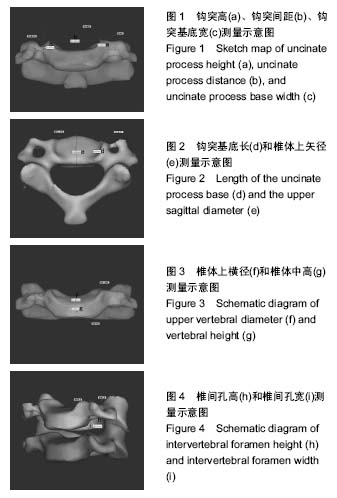
钩椎关节:又称Luschka 关节,是指C3-7 (或C3-T1)下位椎体侧缘钩突内侧缘和上位椎体的下外侧缘斜坡共同组成的4对或5对“类似滑膜关节”的特殊力学结构。钩椎关节周围结构毗邻复杂:后方为脊髓、脊膜支和椎体的血管;后外侧部构成椎间孔的前壁,邻接颈神经根;外侧有椎动静脉和交感神经丛。随年龄增长,椎体钩突常出现骨质增生,可能压迫脊神经或椎血管。
钩突:为颈椎C3-7上外侧缘的骨性突起,位于颈椎椎体后外侧,前方为颈长肌,外侧为横突孔,其内通过椎动静脉及包绕的交感神经丛,后外侧参与构成椎间孔前壁,有颈神经根及根动脉通过,内侧为椎间盘。颈椎钩突的骨质增生在神经根型和椎动脉型颈椎病的发病机制中具有十分重要的地位。
摘要
背景:随着现代生活习惯的改变,使得颈椎病和颈椎相关疾病的发病愈来愈趋于低龄化,基于这一特征,探讨青少年颈椎钩突和椎体间的关系和规律特征,可为颈椎病的早期诊断与预防、治疗提供理论依据。
目的:对青少年颈椎钩突与椎体和椎间孔间相关结构进行相关性分析。
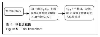
方法:选取无外伤、无神经症状和体征的6-20岁青少年66例,行多排螺旋CT薄层扫描,范围C1-T1,将原始数据以DICOM格式导入三维重建软件进行相关指标测量和后期统计分析。
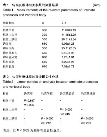
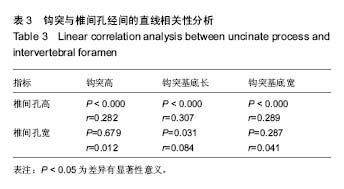
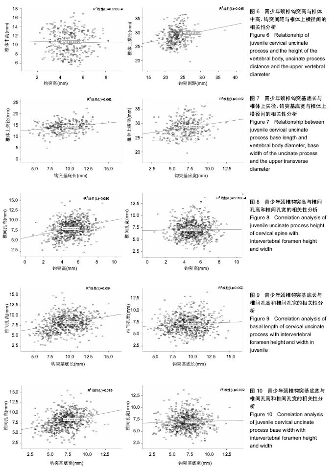
结果与结论:①除钩突高与椎体中高无显著相关性外,钩突间距与椎体上横径、钩突基底宽与椎体上横径、钩突基底长与椎体上矢径之间均存在正相关性(P < 0.05);②除椎间孔宽与钩突高以及椎间孔宽与钩突基底宽间无相关性外,钩突高与椎间孔高、钩突基底长与椎间孔高、钩突基底长与椎间孔宽、钩突基底宽与椎间孔高之间均存在正相关性(P < 0.05);③结果说明,青少年颈椎钩椎关节中钩突与椎体和椎间孔之间存在着一定的相关性,随着年龄的增长、颈椎活动度的增加及脊柱的生长发育,钩突增生、外伤、骨折等因素均可压迫椎间孔内的脊神经从而引起相应的神经根型颈椎病。
中图分类号:
.jpg)