中国组织工程研究 ›› 2022, Vol. 26 ›› Issue (6): 924-928.doi: 10.12307/2022.178

• 脊柱植入物Spinal implants • 上一篇    下一篇

应用体表地形图量化腰椎间盘突出症腰型客观化指标:下腰曲线弹性固定转折点的三维成角

马  超1,王  飞2,刘晓民3,王子昀2,许  奎2,杨文东2,冯  伟2   

  1. 1北京中医药大学,北京市   100029;2空军特色医学中心中西医结合正骨科,北京市   100142;3北京工业大学电子信息与控制工程学院,北京市   100024
  • 收稿日期:2021-04-19 修回日期:2021-04-20 接受日期:2021-05-21 出版日期:2022-02-28 发布日期:2021-12-07
  • 通讯作者: 冯伟,全国名老中医冯天有教授学术经验继承人(新医正骨疗法),主任医师,北京中医药大学硕士生导师,空军特色医学中心中西医结合正骨科,北京市 100142
  • 作者简介:马超,男,1995年生,新疆维吾尔自治区塔城市人,回族,北京中医药大学在读硕士,目前主要从事中西医结合治疗软组织损伤研究。 王飞,男,1975年生,汉族,2016年中国中医科学院毕业,博士,副主任医师,主要从事损伤退变性脊柱疾病的研究。
  • 基金资助:
    空军军医大学课题(2019Z T A03),课题负责人:冯伟

Quantification of the objective index of lumbar disc herniation with body surface topography map: three-dimensional angulation of the elastically fixed turning point of the lower back curve

Ma Chao1, Wang Fei2, Liu Xiaomin3, Wang Ziyun2, Xu Kui2, Yang Wendong2, Feng Wei2   

  1. 1Beijing University of Chinese Medicine, Beijing 100029, China; 2Department of Orthopedics of Integrated Traditional Chinese and Western Medicine, Air Force Special Medical Center, Beijing 100142, China; 3School of Electronic Information and Control Engineering, Beijing University of Technology, Beijing 100024, China
  • Received:2021-04-19 Revised:2021-04-20 Accepted:2021-05-21 Online:2022-02-28 Published:2021-12-07
  • Contact: Feng Wei, Chief physician, Master’s supervisor, Department of Orthopedics of Integrated Traditional Chinese and Western Medicine, Air Force Special Medical Center, Beijing 100142, China
  • About author:Ma Chao, Master candidate, Beijing University of Chinese Medicine, Beijing 100029, China Wang Fei, MD, Associate chief physician, Department of Orthopedics of Integrated Traditional Chinese and Western Medicine, Air Force Special Medical Center, Beijing 100142, China Ma Chao and Wang Fei contributed equally to this article.
  • Supported by:
    Project of Air Force Military Medical University, No. 2019Z T A03 (to FW)

摘要:

文题释义:
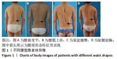
下腰曲线转折点的三维成角:对Ⅲ、Ⅳ型腰椎间盘突出症患者而言,单(多)个椎体发生位移,棘突连线所呈直线中断,成角形成一或多个折线,同时造成脊柱畸形曲线弹性固定,经下腰曲线弹性固定转折点的脊沟曲线、截面曲线在冠状面、矢状面、轴面的投影线与人体竖直线和水平线所形成的夹角。该角的大小决定了两侧椎旁肌形态对称性、髂嵴两侧高低对称性以及人体脊柱旋转的程度。
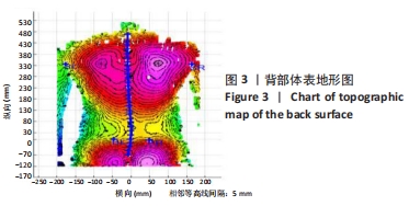
体表地形图:基于双目立体视觉方法,可以产生代表物体表面形状的等高线波纹,随体表形状不同而变形,可用于脊柱变形引发背部表面不对称性的测量,定量分析脊柱侧凸和旋转角度,实现人体背部形态及脊柱非侵入性的数字化和直观再现。

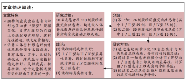
背景:腰椎间盘突出症患者背部形态呈现特征性表现,目前对腰型的判断主要通过望诊观察、采用定性描述,冯天有教授从望诊角度总结出“腰型”四步规律——腰曲变平、腰骶(功能)上移、旋盆翘臀、旋腰挺胸。
目的:测量Ⅲ/Ⅳ型与Ⅰ/Ⅱ型腰椎间盘突出症患者下腰曲线转折点的三维成角,分析通过三维成角判断腰型的可行性。
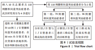
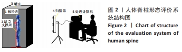
方法:顺次纳入住院腰椎间盘突出症患者110例,其中Ⅰ/Ⅱ腰型70例,Ⅲ/Ⅳ腰型40例。通过人体脊柱形态评价系统采集患者背部体表地形图,应用分析软件测量患者三维成角,初步判断三维成角的稳定性,计算Ⅲ/Ⅳ型与Ⅰ/Ⅱ型患者三维成角的差异性及诊断临界值,并对三维成角的真实性进行初步评价。
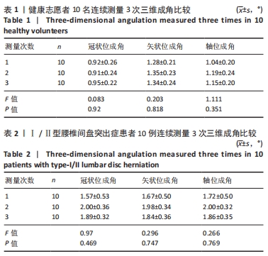
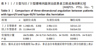
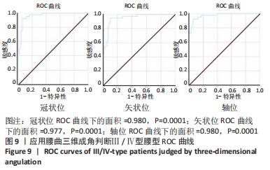
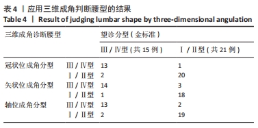
结果与结论:①此次指标稳定性良好,3次三维成角测量数据比较,差异无显著性意义(P > 0.05);②Ⅲ/Ⅳ型与Ⅰ/Ⅱ型患者的三维成角差异极显著:冠状位成角Ⅲ/Ⅳ型平均11.04°,远大于Ⅰ/Ⅱ型(中位数1.24°);矢状位成角Ⅲ/Ⅳ型平均9.02°,远大于Ⅰ/Ⅱ型(平均数2.96°);轴位成角Ⅲ/Ⅳ型平均9.65°,远大于Ⅰ/Ⅱ型(平均数3.01°);③依据ROC曲线值得出Ⅲ/Ⅳ型腰型患者的三维成角(冠状位、矢状位、轴位)的诊断临界值分别为5.43°,4.77°,4.83°。此次三维成角结果真实性可靠,冠状位、矢状位、轴位成角结果显示:灵敏度分别为86.66%,93.33%,86.66%;特异度分别为95.23%,85.72%,90.47%;约登指数分别为0.818,0.790,0.771;总符合率分别为91.66%,88.88%,88.88%;④该研究利用人体背部体表三维地形图技术,分析通过三维成角判断腰型的可行性,发现该指标稳定性好,尤其能够反映旋盆翘臀和旋腰挺胸型的特征,该角度作为一个腰椎间盘突出症腰型分类的关键指标,对于定量描述腰型变化迈出了重要的一步。

https://orcid.org/0000-0003-3665-9133 (马超) 

中国组织工程研究杂志出版内容重点:人工关节;骨植入物;脊柱;骨折;内固定;数字化骨科;组织工程

关键词: 腰椎间盘突出症, 腰型, 体表地形图, 三维成角, 数字化骨科

Abstract: BACKGROUND: The back morphology of patients with lumbar disc herniation presents characteristic manifestations. At present, the judgment of lumbar type is mainly through observation and qualitative description. Professor Feng Tianyou summed up the four steps of “lumbar types” from the perspective of observation: the lumbar curve becomes flat, the lumbosacral upward movement, the rotary basin-buttock, and the rotary waist and chest.  
OBJECTIVE: To measure the three-dimensional angulation at the turning point of low back curve between type-III/IV and type-I/II patients and analyze the feasibility of judging lumbar type by three-dimensional angulation.
METHODS:  A total of 110 hospitalized patients with lumbar intervertebral disc herniation were enrolled sequentially, including 70 cases of type-I/II patients and 40 cases of type-III/IV patients. The topographic map of the patient’s back surface was collected through the human spine morphology evaluation system. The analysis software was used to measure the patient’s three-dimensional angulation. The stability of three-dimensional angulation was preliminarily determined. The difference and diagnostic critical value of three-dimensional angulation between type-III/IV and type-I/II patients were calculated, and the authenticity of 3D angle formation was evaluated.  
RESULTS AND CONCLUSION: (1) The index was stable, and there was no statistical significance in the comparison of the three times for three-dimensional angulation measurement data (P > 0.05). (2) There was significant difference in three-dimensional angulation between type-III/IV and type-I/II patients. Coronal angulation of type-III/IV was 11.04° on average, which was much larger than that of type-I/II (median 1.24°). Sagittal angulation of type-III/IV was 9.02° on average, which was much larger than that of type-I/II (mean 2.96°). Axial angulation of type-III/IV was 9.65° on average, which was much larger than that of type-I/II (mean 3.01°). (3) According to the ROC curve, the diagnostic critical value of three-dimensional angulation (coronal, sagittal, and axial) in III/IV-type patients were 5.43°, 4.77°, and 4.83°, respectively. The authenticity of the three-dimensional angulation results was reliable. The coronal, sagittal, and axial angulation results showed that the sensitivity was 86.66%, 93.33%, and 86.66%; the specificity was 95.23%, 85.72%, and 90.47%. The Youden index was 0.818, 0.790, and 0.771. The total compliance rate was 91.66%, 88.88%, and 88.88%. (4) In this study, three-dimensional topographic map technology of human back surface was used to analyze the feasibility of judging the lumbar type by three-dimensional angulation. It was found that the index had good stability, especially could reflect the characteristics of the rotary basin-buttock, and the rotary waist and chest.

Key words: lumbar disc herniation, lumbar types, body surface topographic map, three-dimensional angulation, digital orthopedics

中图分类号: