中国组织工程研究 ›› 2016, Vol. 20 ›› Issue (29): 4297-4302.doi: 10.3969/j.issn.2095-4344.2016.29.006
• 软骨组织构建 cartilage tissue construction • 上一篇 下一篇
AMP活化蛋白激酶在终板软骨细胞体外传代培养中的表达及意义
赵泉来,郑 权,徐宏光,沈 祥,王 弘,刘 平,王凌挺,杨晓明,陈学武,张 玙,李逸峰,俞宏星
- 皖南医学院第一附属医院弋矶山医院脊柱外科,安徽省芜湖市 241001
Expression of AMP-activated protein kinase in subcultured rat endplate chondrocytes
Zhao Quan-lai, Zheng Quan, Xu Hong-guang, Shen Xiang, Wang Hong, Liu Ping, Wang Ling-ting, Yang Xiao-ming, Chen Xue-wu, Zhang Yu, Li Yi-feng, Yu Hong-xing
- Department of Spine Surgery, Yijishan Hosptial, Wannan Medical College, Wuhu 241001, Anhui Province, China
摘要:
文章快速阅读:
.jpg) 文题释义:
终板软骨:终板软骨为附着于椎体上下缘的一层透明软骨,它能够将椎体末端血管中的营养物质通过渗透作用运输至髓核和内层纤维环,是椎间盘主要的营养途径,终板营养途径阻断,可致椎间盘内Ⅱ型胶原、蛋白聚糖等表达减少,椎间盘发生退变。
AMP活化蛋白激酶:AMP活化蛋白激酶(AMPK)是真核生物中一种重要的蛋白激酶,其在各种与代谢相关的器官中均有表达,能够协调调节细胞的合成代谢和分解代谢,因此被称为细胞能量调节器,近年的研究表明AMP活化蛋白激酶活性与软骨的形成和降解也有重要的关系。
文题释义:
终板软骨:终板软骨为附着于椎体上下缘的一层透明软骨,它能够将椎体末端血管中的营养物质通过渗透作用运输至髓核和内层纤维环,是椎间盘主要的营养途径,终板营养途径阻断,可致椎间盘内Ⅱ型胶原、蛋白聚糖等表达减少,椎间盘发生退变。
AMP活化蛋白激酶:AMP活化蛋白激酶(AMPK)是真核生物中一种重要的蛋白激酶,其在各种与代谢相关的器官中均有表达,能够协调调节细胞的合成代谢和分解代谢,因此被称为细胞能量调节器,近年的研究表明AMP活化蛋白激酶活性与软骨的形成和降解也有重要的关系。
文题释义:
终板软骨:终板软骨为附着于椎体上下缘的一层透明软骨,它能够将椎体末端血管中的营养物质通过渗透作用运输至髓核和内层纤维环,是椎间盘主要的营养途径,终板营养途径阻断,可致椎间盘内Ⅱ型胶原、蛋白聚糖等表达减少,椎间盘发生退变。
AMP活化蛋白激酶:AMP活化蛋白激酶(AMPK)是真核生物中一种重要的蛋白激酶,其在各种与代谢相关的器官中均有表达,能够协调调节细胞的合成代谢和分解代谢,因此被称为细胞能量调节器,近年的研究表明AMP活化蛋白激酶活性与软骨的形成和降解也有重要的关系。摘要
背景:终板软骨功能障碍是椎间盘退变的始动因素,AMPK在软骨的形成和降解过程中也具有相应的调节作用。
目的:观察AMP活化蛋白激酶(AMPK)在终板软骨细胞体外自然传代退变模型中的作用。
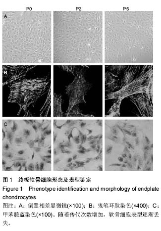
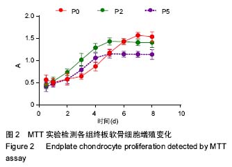
方法:分离大鼠腰椎终板软骨细胞,体外自然传代培养,取原代细胞(P0)、第2代细胞(P2)及第5代细胞(P5),倒置显微镜及细胞骨架染色观察终板软骨细胞形态的变化,甲苯胺蓝染色观察终板软骨细胞表型的改变;MTT法检测3组细胞的增殖能力;通过Real time-PCR评价3组终板软骨细胞中软骨标志基因(Ⅱ型胶原、蛋白多糖、SOX-9)以及基质金属蛋白酶3和13的表达变化,Western blot检测终板软骨细胞中AMPK蛋白磷酸化的改变。
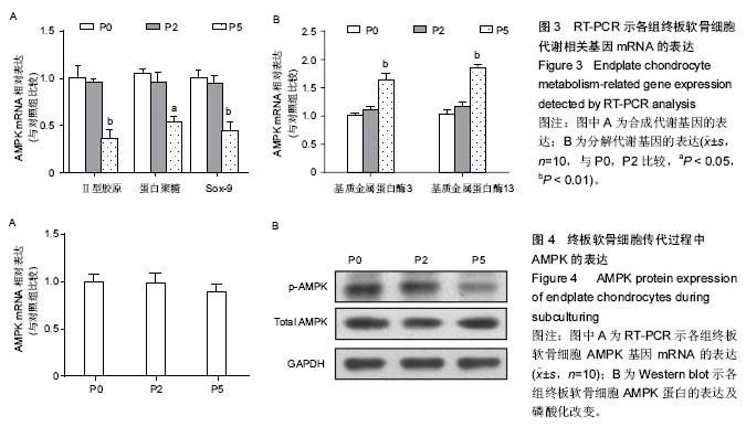
结果与结论:①随着传代次数的增加终板软骨细胞形态发生改变,增殖能力降低,软骨细胞表型逐渐丢失;②P5代终板软骨细胞中Ⅱ型胶原、蛋白聚糖及SOX-9基因表达明显下调(P < 0.05或0.01),而基质金属蛋白酶3, 基质金属蛋白酶13基因表达较P0代及P2代均明显增高(P < 0.01);③Western blot 结果显示P5代终板软骨细胞中AMPK蛋白磷酸化水平明显降低。提示:AMPK磷酸化的水平与终板软骨细胞体外自然传代过程中软骨细胞的退变密切相关,调控AMPK活性有可能减轻或阻止终板软骨及椎间盘的退变。
中国组织工程研究杂志出版内容重点:组织构建;骨细胞;软骨细胞;细胞培养;成纤维细胞;血管内皮细胞;骨质疏松;组织工程
ORCID: 0000-0002-5619-8082(赵泉来)
中图分类号:
.jpg) 文题释义:
终板软骨:终板软骨为附着于椎体上下缘的一层透明软骨,它能够将椎体末端血管中的营养物质通过渗透作用运输至髓核和内层纤维环,是椎间盘主要的营养途径,终板营养途径阻断,可致椎间盘内Ⅱ型胶原、蛋白聚糖等表达减少,椎间盘发生退变。
AMP活化蛋白激酶:AMP活化蛋白激酶(AMPK)是真核生物中一种重要的蛋白激酶,其在各种与代谢相关的器官中均有表达,能够协调调节细胞的合成代谢和分解代谢,因此被称为细胞能量调节器,近年的研究表明AMP活化蛋白激酶活性与软骨的形成和降解也有重要的关系。
文题释义:
终板软骨:终板软骨为附着于椎体上下缘的一层透明软骨,它能够将椎体末端血管中的营养物质通过渗透作用运输至髓核和内层纤维环,是椎间盘主要的营养途径,终板营养途径阻断,可致椎间盘内Ⅱ型胶原、蛋白聚糖等表达减少,椎间盘发生退变。
AMP活化蛋白激酶:AMP活化蛋白激酶(AMPK)是真核生物中一种重要的蛋白激酶,其在各种与代谢相关的器官中均有表达,能够协调调节细胞的合成代谢和分解代谢,因此被称为细胞能量调节器,近年的研究表明AMP活化蛋白激酶活性与软骨的形成和降解也有重要的关系。