中国组织工程研究 ›› 2015, Vol. 19 ›› Issue (49): 8015-8020.doi: 10.3969/j.issn.2095-4344.2015.49.025

• 相关因子与动物模型 related factors in animal models • 上一篇    下一篇

角膜炎模型大鼠白细胞介素10及环氧酶2的表达

陆 霞1,潘旭斌1,谢玉涛2,季秋娣3   

  1. 无锡市第四人民医院,1眼科,2手术室,3皮肤科,江苏省无锡市 214062
  • 收稿日期:2015-09-23 出版日期:2015-11-30 发布日期:2015-11-30
  • 作者简介:陆霞,女,1966年生,安徽省淮南市人,汉族,2001年南京医科大学毕业,主管护师,主要从事眼科相关工作。

Expression of interleukin-10 and cyclooxygenase 2 in keratitis rat models

Lu Xia1, Pan Xu-bin1, Xie Yu-tao2, Ji Qiu-di3   

  1. 1Department of Ophthalmology, 2Operating Room, 3Department of Dermatology, the Fourth People’s Hospital of Wuxi, Wuxi 214062, Jiangsu Province, China
  • Received:2015-09-23 Online:2015-11-30 Published:2015-11-30
  • About author:Lu Xia, Nurse-in-charge, Department of Ophthalmology, the Fourth People’s Hospital of Wuxi, Wuxi 214062, Jiangsu Province, China

摘要:

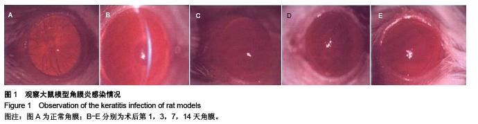
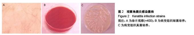
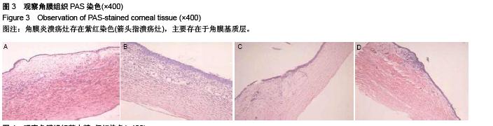
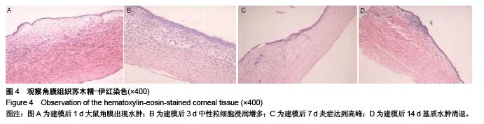
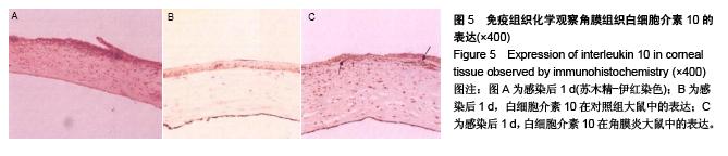
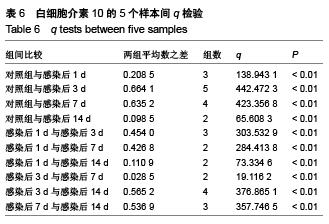
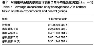
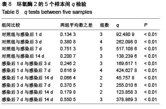
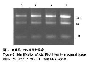
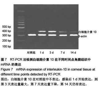
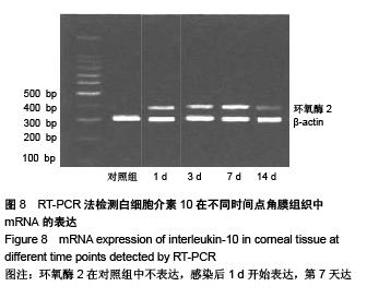
背景:研究白细胞介素10、环氧酶2与模型大鼠角膜炎发生发展的关系,对角膜炎的治疗有重大意义。
目的:检测白细胞介素10及环氧酶2在大鼠模型角膜炎中的表达情况,分析白细胞介素10、环氧酶2与模型大鼠角膜炎发生发展的关系。
方法:随机选取26只健康大鼠,不限雌雄,大鼠左眼选为实验组,采用角膜表面镜片术法建立角膜炎感染模型;右眼作为正常对照组。建模后第1,3,7及14天摘取角膜组织,观察大鼠左右眼角膜组织病理学变化,并采用免疫组织化学及反转录聚合酶链式反应(RT-PCR)法,检测白细胞介素10及环氧酶2在不同程度角膜炎的角膜组织中的表达情况。
结果与结论:①组织病理学观察显示角膜炎病灶有严重组织坏死及中性粒细胞浸润,PAS染色结果显示菌丝主要存在于角膜基质层。②白细胞介素10在对照组大鼠角膜中基本不表达,感染角膜炎后,白细胞介素10的表达先上升后下降,各时间点有显著性差异(P < 0.01)。③环氧酶2在对照组大鼠角膜中基本不表达,感染角膜炎后,环氧酶2的表达逐渐升高,第14天降低,各时间点有显著性差异(P < 0.01)。PAS染色及病变组织培养实验证明建立大鼠模型角膜炎成功。结果显示白细胞介素10在角膜炎病变后期有表达,推测其可能参与角膜炎后期的组织损伤修复;环氧酶2的表达位于角膜炎组织,是角膜炎病变的敏感炎症因子。
 

关键词: 实验动物模型, 基因病毒载体及相关因子动物模型, 角膜炎, 白细胞介素10, 环氧酶2

Abstract:

BACKGROUND: Investigating the relationship between interleukin-10, cyclooxygenase-2 expression levels and the occurrence and development of keratitis is of great significance for the treatment of keratitis.
OBJECTIVE: To detect the expression of interleukin-10 and cyclooxygenase 2 in keratitis rat models and analyze the relationship between interleukin-10, cyclooxygenase-2 expression and the occurrence and development of keratitis in rats.
METHODS: Twenty-six healthy rats of either gender were randomly selected to establish keratitis rat models. The left eyes of rats were chosen as experimental group to establish keratitis models using epikeratophakia. The right eyes of rats were set as control group. At the 1th, 3rd, 7th and 14th days after modeling, the pathological changes of rat corneal tissue were observed in both eyes. The expression of interleukin-10 and cyclooxygenase 2 in the corneal tissue with different degrees of keratitis was determined using immunohistochemistry and reverse transcription-polymerase chain reaction (RT-PCR).      
RESULTS AND CONCLUSION: Histopathological observation showed that there were severe tissue necrosis and neutrophil infiltration in keratitis foci. PAS staining showed that the mycelium was mainly present in the corneal stroma. In the control group, the expression of interleukin-10 was hardly detected in rat cornea. After keratitis infection, the expression of interleukin 10 increased at first and then decreased, and there was significant difference between different time points (P < 0.01). In the control group, the expression of cyclooxygenase-2 was hardly detected in rat cornea. After keratitis infection, the expression of cyclooxygenase 2 was increased gradually and then decreased at the 14th day. There was significant difference between different time points (P < 
0.01). PAS staining and pathologic tissue culture experiment suggest that rat models of keratitis were successfully established. These results show that the expression of interleukin-10 appeared in the late period of keratitis lesions, which indicate that interleukin-10 may be involved in the tissue damage repair in the late period of keratitis. The expression of cyclooxygenase-2 locates in the keratitis tissue and cyclooxygenase-2 is a sensitive inflammatory cytokine of keratitis.  


中图分类号: