中国组织工程研究 ›› 2015, Vol. 19 ›› Issue (37): 5943-5949.doi: 10.3969/j.issn.2095-4344.2015.37.008
• 软骨组织构建 cartilage tissue construction • 上一篇 下一篇
盘状半月板损伤修复:关节镜下缝合成形结合玻璃酸钠注射
唐 进,吉 明,廖乙名,程智勇,陈 林,占方彪,杨 涛
- 三峡中心医院骨科,重庆市 404000
-
出版日期:2015-09-10发布日期:2015-09-10 -
通讯作者:杨涛,主治医师,三峡中心医院骨科,重庆市 404000 -
作者简介:唐进,男,1984年生,重庆市人,汉族,2011年重庆医科大学毕业,硕士,主治医师,主要从事骨关节外科方面的研究。
Arthroscopic all-inside suture repair combined with sodium hyaluronate injection for discoid meniscus injury
Tang Jin, Ji Ming, Liao Yi-ming, Cheng Zhi-yong, Chen Lin, Zhan Fang-biao, Yang Tao
- Department of Orthopedics, Chongqing Three Gorges Central Hospital, Chongqing 404000, China
-
Online:2015-09-10Published:2015-09-10 -
Contact:Yang Tao, Attending physician, Department of Orthopedics, Chongqing Three Gorges Central Hospital, Chongqing 404000, China -
About author:Tang Jin, Master, Attending physician, Department of Orthopedics, Chongqing Three Gorges Central Hospital, Chongqing 404000, China
摘要:
背景:研究表明,关节镜下缝合成形治疗膝关节半月板损伤临床效果满意,而结合玻璃酸钠对促进软骨修复有重要作用。 目的:分析关节镜下缝合成形结合玻璃酸钠注射促进膝关节盘状半月板损伤的软骨修复。 方法:关节镜下缝合成形结合玻璃酸钠注射修复盘状半月板损伤22例,术中证实均为附着缘松弛或纵裂。采用关节镜下缝合成形术结合玻璃酸钠注射治疗,继而进行有效的康复计划。 结果与结论:22例患者治疗全部成功,无并发症。患者均获得18个月以上随访,术后疗效根据Ikenchi评分方法:优11例,良8例,可3例,优良率86%。膝关节镜下盘状半月板缝合成形可以最大程度地保留半月板解剖结构,使其发挥有效功能且损伤小,关节修复后配和关节腔注射玻璃酸钠及正规的康复训练并发症少,可有效促进半月板关节软骨修复。
中国组织工程研究杂志出版内容重点:组织构建;骨细胞;软骨细胞;细胞培养;成纤维细胞;血管内皮细胞;骨质疏松;组织工程
中图分类号:
引用本文
唐 进,吉 明,廖乙名,程智勇,陈 林,占方彪,杨 涛. 盘状半月板损伤修复:关节镜下缝合成形结合玻璃酸钠注射[J]. 中国组织工程研究, 2015, 19(37): 5943-5949.
Tang Jin, Ji Ming, Liao Yi-ming, Cheng Zhi-yong, Chen Lin, Zhan Fang-biao, Yang Tao. Arthroscopic all-inside suture repair combined with sodium hyaluronate injection for discoid meniscus injury [J]. Chinese Journal of Tissue Engineering Research, 2015, 19(37): 5943-5949.
Quantitative analysis of participants
| [1] Fu D, Guo L, Yang L, et al. Discoid lateral meniscus tears and concomitant articular cartilage lesions in the knee. Arthroscopy. 2014;30(3):311-318. [2] Ahn J H, Kim K I, Wang J H, et al. Long-term results of arthroscopic reshaping for symptomatic discoid lateral meniscus in children. Arthroscopy. 2015;31(5):867-873. [3] Dai Z,Chen ZW. Research in meniscus root tear. Zhongguo Jiaoxing Waike Zazhi. 2014;22(8):715-718. [4] Huang CZ, Fan WJ, Chen ZW, et al. Application of menisci reformation and repair in the treatment of the discoid meniscus injuries. Zhongguo Gushang. 2010;23 (6): 409-412. [5] lkeuchi H. Arthroscopic treatment of the discoid lateral meniscus technique and long term results. Clin Orthop Relat Res. 1982;(167):19-28. [6] Liu YJ, Wang Y, Wang LE. Applied Arthroscopic Surgery. Beijing: People’s Military Medical Press.2006: 47-50. [7] Chung JY, Roh JH, Kim JH, et al. Bilateral occurrence and morphologic analysis of complete discoid lateral meniscus. Yonsei Med J. 2015;56(3):753-759. [8] Ding J, Zhao J, He Y, et al. Risk factors for articular cartilage lesions in symptomatic discoid lateral meniscus. Arthroscopy. 2009;25(12):1423-1426. [9] Ao YF. Arthroscopic Knee Surgery. Beijing: Peking University Medical Press. 2004:120. [10] Roberts SB, Beattie N, Brown G, et al. Interventions for treating injuries of the medial ligaments of the knee. The Cochrane Library. 2014. [11] Forst R. Bedeutung der orthopädischen Krankheitsbilder in der Geriatrie[J]. Mobilität im Alter. Herausforderungen für Orthopädie und Unfallchirurgie. Deutscher Ärzte-Verlag GmbH Köln. 2006. [12] Shah AM, Mann DL. In search of new therapeutic targets and strategies for heart failure: recent advances in basic science. The Lancet. 2011;378(9792):704-712. [13] Holzmeister J, Leclercq C. Implantable cardioverter defibrillators and cardiac resynchronisation therapy. The Lancet. 2011;378(9792):722-730.[14] Dou XL,Duan XQ,Xia L,et al.Osteoarthritis: study and progress in articular cartilage degeneration.Zhongguo Zuzhi Gongcheng Yanjiu. 2011;15(20):3763-3766. [15] Wang ZB, Xu YL, Liao WM, et al. Cartilage surface of early osteoarthritis in rats induced by papain under scanning electron microscope. Zhongguo Zuzhi Gongcheng Yanjiu. 2014;18(2):177-182. [16] Lee DH, Kim TH, Kim JM, et al. Resdts of subtotal/total or partial meniscectomy for discoid lateral meniscus in children. Arthroscopy. 2009;25(5):496-503. [17] Ahn JH, Lee SH, Yoo JC, et al. Bilateral discoid lateral meniscus in knees: evaluation of the contralateral knee in patients with symptomatic discoid lateral meniscus. Arthroscopy. 2010;26(10):1348-1356. [18] Ikeuchi H. Arthroscopie treatment of the discoid lateral meniscus:technique and long termresults. Clin Orthop. 1998;167:19-28. [19] Wasser L, Knörr J, Accadbled F, et al. Arthroscopic treatment of discoid meniscus in children: clinical and MRI results. Orthop Traumatol Surg Res. 2011;97(3):297-303. [20] Adachi N, Ochi M, Uchio Y, et al. Torn discoid lateral meniscus treated using partial central meniscectomy and suture of the peripheral tear. Arthroscopy. 2004;20(5):536-542. [21] Kim JM, Bin SI. Meniscal allugraft transplantation after total meniscectomy of torn discoid lateral meniscus. Arthroscopy. 2006;22(12):1344-1350. [22] Hommen JP, Applegate GR, Del Pizzo W. Meniscus allograft transplantation:ten-year results of cryopreserved allografts. Arthroscopy. 2007;23(4):388-393. [23] Hayashi LK, Yamaga H, Ida K, et al. Arthroscopic meniscectomy for discoid lateral meniscus in children. J Bone Joint Surg Am. 1988;70(10):1495-1500. [24] Mayer-Wagner S, von Liebe A, Horng A, et al. Discoid lateral meniscus in children: magnetic resonance imaging after arthroscopic resection. Knee Surgery. Sports Traumatology, Arthroscopy. 2011;19(11):1920-1924. [25] Miller MD, Ritchie JR, Gomez BA, et al. Meniscal repair: an experimental study in the goat. Am J Sports Med. 1995; 23(1):124-128. [26] Henning CE. Arthroscopic repair of meniscus tears. Orthopedics. 1983;6(9):1130-1132. [27] Chen S, Fu PL, Cong RJ, et al. Research progress of meniscus tissue engineering. Zhonggou Gu yu Guanjie Zazhi. 2013;2(12):694-698. [28] Zhong WJ, Wang YB, Zhang YD, et al. Experimental study of effect of meniscus suture on meniscus healing. Zhongguo Xiufu Zhongjian Waike Zazhi. 2000;14(21): 77-79. [29] Li X, Zhong J, Hong L, et al. Hybrid suture technique for repairing bucket-handle tear of the lateral meniscus. Zhonghua Guke Zazhi. 2012;32(2):101-105. [30] He RZ, Wu S, Cao X, et al. Therapeutic Effect of Early Rehabilitative Training after Arthroscopic Anterior Cruciate Ligament Reconstruction. Xiandai Shengwu Yixue Jinzhan. 2012;12(7):1274-1276,1269. |
| [1] | 张同同, 王中华, 文 杰, 宋玉鑫, 刘 林. 3D打印模型在颈椎肿瘤手术切除与重建中的应用[J]. 中国组织工程研究, 2021, 25(9): 1335-1339. |
| [2] | 韦 玮, 李 剑, 黄林海, 兰敏东, 卢显威, 黄绍东. 全膝或全髋关节置换后老年人首次活动时跌倒恐惧的影响因素[J]. 中国组织工程研究, 2021, 25(9): 1351-1355. |
| [3] | 王金军, 邓增发, 刘 康, 何智勇, 余新平, 梁建基, 李 晨, 郭洲洋. 全膝关节置换静脉滴注氨甲环酸联合含氨甲环酸鸡尾酒局部应用的止血效果及安全性[J]. 中国组织工程研究, 2021, 25(9): 1356-1361. |
| [4] | 肖国庆, 刘选泽, 严钰皓, 钟喜红. 后交叉韧带替代型假体全膝关节置换术后屈曲受限的影响因素[J]. 中国组织工程研究, 2021, 25(9): 1362-1367. |
| [5] | 黄泽晓, 杨 妹, 林诗炜, 何和与. 血清n-3多不饱和脂肪酸水平与全膝关节置换早期股四头肌肌力变化的相关性[J]. 中国组织工程研究, 2021, 25(9): 1375-1380. |
| [6] | 张尚普, 鞠晓东, 宋恒义, 董 智, 王 晨, 孙国栋. 关节镜下带线锚钉缝线桥缝合固定治疗肩锁关节脱位[J]. 中国组织工程研究, 2021, 25(9): 1417-1422. |
| [7] | 黄登承, 王志科, 曹学伟. 体外冲击波疗法治疗中老年膝骨关节炎短期疗效对比的荟萃分析[J]. 中国组织工程研究, 2021, 25(9): 1471-1476. |
| [8] | 曾燕华, 郝延磊. 许旺细胞体外培养及纯化的系统性综述[J]. 中国组织工程研究, 2021, 25(7): 1135-1141. |
| [9] | 仲鹤鹤, 孙鹏鹏, 桑 鹏, 吴术红, 刘 毅. 模拟重建膝关节后外侧复合体核心韧带后膝关节稳定性评估[J]. 中国组织工程研究, 2021, 25(6): 821-825. |
| [10] | 赵中溢, 李勇阵, 陈 峰, 季爱玉. 同期双侧全膝关节置换和单髁置换治疗创伤性关节炎的比较[J]. 中国组织工程研究, 2021, 25(6): 854-859. |
| [11] | 刘少华, 周观明, 陈希聪, 肖可明, 蔡 剑, 刘效仿. 前交叉韧带缺陷对固定平台单髁置换后中期疗效的影响[J]. 中国组织工程研究, 2021, 25(6): 860-865. |
| [12] | 张念军, 陈 茹. 全膝关节置换过程中鸡尾酒疗法联合股神经阻滞的镇痛效果[J]. 中国组织工程研究, 2021, 25(6): 866-872. |
| [13] | 袁 俊, 杨家福. 局部氨甲环酸浸润在非骨水泥全膝关节置换过程中止血效果的评价[J]. 中国组织工程研究, 2021, 25(6): 873-877. |
| [14] | 张 磊, 马 丽, 扶世杰, 周 鑫, 喻 林, 郭晓光. 肩关节镜下双排锚钉固定治疗肩关节前脱位伴肱骨大结节撕脱骨折[J]. 中国组织工程研究, 2021, 25(6): 895-900. |
| [15] | 黄登承, 王志科, 曹学伟.
全膝关节置换中静脉和局部联合或单一应用氨甲环酸:一项随机对照试验的荟萃分析
[J]. 中国组织工程研究, 2021, 25(6): 948-956. |
Design
A before-and-after study.
Time and setting
The experiment was completed in the Department of Orthopedics, Chongqing Three Gorges Central Hospital from January 2011 to December 2014.
Subjects
From January 2011 to March 2013, 22 cases of lateral discoid meniscus undergoing arthroscopic surgery were enrolled, 9 males and 13 females, aged 21-46 years (mean 29.8 years). All the cases were confirmed to have attached edge relaxation or longitudinal crack in operation. The course of disease was 30 days to 48 months. Clinical manifestations included a history of trauma in 18 cases, pain in 21 cases, joint locking in 19 cases, joint snapping in 19 cases, quadriceps atrophy in 14 cases, positive McMurray test in 17 cases. Anteroposterior X-ray examination of the knee showed lateral joint space was widened in 17 cases.
Inclusion criteria: In addition to the clinical symptoms and signs, the following aspects are included: (1) MRI performance: the meniscus is enlarged, widened and thickened, the major performance is that three consecutive sagittal plane layers or above exhibit the anterior and posterior horns of the meniscus is connected in a “bowtie” like manner, and the posterior horn is thickened obviously; three consecutive coronal plane layers or above display the meniscus width was more than half of the tibial plateau. (2) The disease is confirmed during arthroscopic surgery. (3) Attached edge relaxation or longitudinal crack is confirmed during surgery.
Exclusion criteria: (1) local skin ulceration, infection, bullous and other skin diseases. (2) Normal meniscus injury. (3) Discoid meniscus injury associated with complete-type discoid meniscus injury with crack damage. (4) Patients with hearing and speaking disorder or cannot cooperate with medical treatment.
Materials: Sodium hyaluronate (Kunming Baker Norton Pharmaceutical Co., Ltd., China) is a polysaccharide polymer biomaterial composed of N-acetyl glucuronide that is repeatedly alternated. Sodium hyaluronate as a main component of synovial fluid is one component of the cartilage matrix, which plays an intra-articular lubrication role, cover and protect the articular cartilage, improve joint contractures, inhibit surface changes due to cartilage degeneration, improve pathological synovial fluid, and increase drop-slip function.
Methods
Arthroscopic suture combined with sodium hyaluronate treatment
Under spinal or epidural anesthesia, the patient was in the supine position, the upper third of the thigh was subjected to conventional hemostasis (not exceeding 90 minutes), and the knee joint was in 90° flexion or “4” position, which can be alternate. The articular cavity was explored via conventional anteromedial and anterolateral approaches in order: suprapatellar bursa, patellofemoral joint, medial compartment, intercondylar fossa and lateral compartment, to carefully observe the shape of discoid lateral meniscus, understand the type and scope of damage, and determine the surgical method. In this study, all cases were confirmed to have incomplete discoid meniscus or mild complete-type discoid meniscus with attached edge relaxation or longitudinal crack, but not to have complete-type discoid meniscus with crack damage. The center of discoid meniscus was removed by medical pliers with edge-preserving width of 6-8 mm, and then the edge of the meniscus was trimmed using a planing device and plasma knife so as to form a ramp similar to the normal meniscus. For attached edge relaxation or longitudinal crack, the inside-out suture repair was preferred. Under the arthroscopy, the edge of injured meniscus was freshed, and sutured with an interval of 5 mm at different angles. Needle and stitching thread passed through the tear edges, so that the one end of the thread was out of the skin, and the other end was outside the entrance of the knee instrument. The same suturing method was run, the one end of the thread pass through the outer skin via the meniscus followed by tightening the two ends of the thread, and the tear hole was folded under arthroscope. Then, a skin incision was made at the exit, and the suture knot was placed under the skin, thus completing a suture. Before the end of surgery, knee hyperextension, flexion and Maxwell test were done to ensure that snapping signs disappeared; then, the wound was sutured and suture wounds, sodium hyaluronate (25 mg) was injected intra-articularly followed by pressure bandaging of the knee. After operation, sodium hyaluronate (25 mg) was injected once a week, totally for 4 weeks.
Postoperative treatment and rehabilitation exercises
The knee joint was fixed in the extension position using a brace. Within 2-3 days after operation, the knee joint was covered with elastic bandage followed by conventional ice compress, and under limit load, the patients began to do muscular training, isometric contraction of the quadriceps and ankle pump exercise until the quadriceps and hamstrings restored the muscle strength. Two or three days later, the patients could walk with crutches; meanwhile, the patients underwent non-weight-bearing
exercise at a movement range of 0°-90°. The joint activities gradually restored after 3 weeks. At 4 weeks after operation, the brace was removed and the patient could walk with crutches under partial weight-bearing, and walk with no crutches under full weight bearing at 8 weeks after operation. Quadriceps isometric exercise was carried out at early stage; within 6 months after operation, sudden stop and squat of the knee and rotational movement of the knee relying on the affected limb (steering run, flashback) should be forbidden.
The evaluation standard
According to Ikeuchi’s method[5], the assessment of efficacy is as follows: excellent for normal joint movement, no snapping, no pain; good for mild pain and no snapping during exercise with normal range of motion; fair for mild pain and sometimes snapping during movement with limited range of motion; poor for pain and snapping during movement with limitation of motion.
Main outcome measures
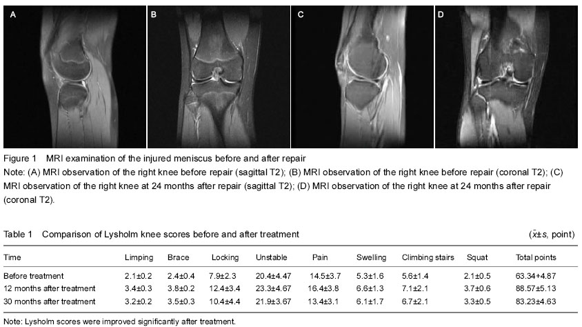
Ikeuchi scores after repair and Lysholm scores before and after repair.
Statistical analysis
Measurement data were expressed as mean±SD and analyzed using SPSS 17.0 statistical software. Paired t-test was used for comparison of Lysholm scores before and after repair. A value of P < 0.05 was considered significant.
1 研究表明,关节镜下缝合成形治疗膝关节半月板损伤临床效果满意,而结合玻璃酸钠对促进软骨修复有重要作用。试验针对盘状半月板附着缘松弛或破裂,在关节镜1下采用缝合、成形并结合玻璃酸钠治疗。从Ikeuchi评分,Lysholm评分等疗效评分方面探讨了关节镜下缝合成形结合玻璃酸钠治疗盘状半月板损伤的临床疗效。
2 试验结果表明,膝关节镜下盘状半月板缝合成形可以最大程度地保留半月板解剖结构,使其发挥有效功能,损伤小,关节修复后配和关节腔注射玻璃酸钠治及正规的康复训练疗,恢复快,并发症少,可获得良好疗效,本组临床疗效满意。
正盘状半月板即盘状软骨,是先天性半月板形态异常,形成大而肥厚的类似圆盘的畸形软骨,多见于膝关节外侧。本病多发生于青壮年,男性发病多于女性,大约(2~7)∶关节镜手术是一种集诊断及治疗于一体的微创手术方法,关节镜下行半月板成形术治疗半月板损伤具有手术创伤小、视野清晰及术后康复快等特点。
| 阅读次数 | ||||||
|
全文 |
|
|||||
|
摘要 |
|
|||||