中国组织工程研究 ›› 2015, Vol. 19 ›› Issue (30): 4800-4804.doi: 10.3969/j.issn.2095-4344.2015.30.009

• 组织工程口腔材料 tissue-engineered oral materials • 上一篇    下一篇

60Co放射及微量元素锌对钛种植体骨融合的影响

陈海军1,张安生2,朱孝春3,钱奇春1,于淑湘1,杨博程1,张静波1,南福清1,林均舟1   

  1. 1解放军第九十七医院口腔科,江苏省徐州市 221004; 2解放军73087部队医院,江苏省徐州市 221011; 3解放军73068部队卫生队,江苏省徐州市 221000
  • 出版日期:2015-07-16 发布日期:2015-07-16
  • 通讯作者: 张安生,博士,主治医师,解放军73087部队医院,江苏省徐州市 221011
  • 作者简介:陈海军,男,1964年生,安徽省萧县人,汉族,1996年解放军第四军医大学口腔医学院毕业,硕士,主任医师,主要从事口腔种植体骨界面力学分析的研究。

Effect of 60Co irradiation and trace element zinc on implant-bone fusion

Chen Hai-jun1, Zhang An-sheng2, Zhu Xiao-chun3, Qian Qi-chun1, Yu Shu-xiang1, Yang Bo-cheng1, Zhang Jing-bo1, Nan Fu-qing1, Lin Jun-zhou1   

  1. 1Department of Stomatology, the 97th Hospital of Chinese PLA, Xuzhou 221004, Jiangsu Province, China; 2Hospital of the 73087 Troop of Chinese PLA, Xuzhou 221011, Jiangsu Province, China; 3Health Team of the 73068 Troop of Chinese PLA, Xuzhou 221000, Jiangsu Province, China
  • Online:2015-07-16 Published:2015-07-16
  • Contact: Zhang An-sheng, M.D., Attending physician, Hospital of the 73087 Troop of Chinese PLA, Xuzhou 221011, Jiangsu Province, China
  • About author:Chen Hai-jun, Master, Chief physician, Department of Stomatology, the 97th Hospital of Chinese PLA, Xuzhou 221004, Jiangsu Province, China

摘要:

背景:前期实验证实补锌可以加速种植体的骨融合。

目的:观察60Co放射及微量元素锌对种植体骨融合的影响。
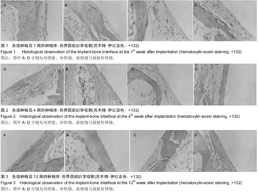
方法:取成年雄性家兔36只,双侧胫骨近心端各植入1枚钛种植体,建立钛种植体动物模型,随机均分为4组,补锌组种植后24 h肌注10 g/L硫酸锌4 mg/kg,1次/d;对照组种植后24 h肌注9 g/L生理盐水4 mg/kg,1次/d;放射组于种植后第2,4,6天给予双侧胫骨近心段60Co照射,15 Gy/d,同时种植后24 h肌注9 g/L生理盐水4 mg/kg,1次/d;放射补锌组于种植后第2,4,6天给予双侧胫骨近心段60Co照射,15 Gy/d,同时种植后24 h肌注10 g/L硫酸锌4 mg/kg,1次/d。种植后1,4,12周利用显微镜观察种植体骨界面组织形态。
结果与结论:在相同时间段内,与其他各组相比,放射组种植体周围成纤维细胞数量较多,纤维性结合较多,而骨性组织特别是成熟骨组织形成较少。在相同时间段内,与其他各组相比,补锌组种植体表面成骨细胞数量更多,骨性结合更多,在第4周时即可见成熟骨组织出现,说明补锌能在种植后早期(1-4周)即形成骨性愈合。放射补锌组在第4,12周时也较放射组产生更多骨性结合,说明即使在60Co照射后,微量元素锌的补充仍可以促进种植体-骨融合。提示60Co照射会延缓种植体骨愈合,放射治疗后适量补锌能够减轻其对种植体骨融合的不利影响。

中国组织工程研究杂志出版内容重点:生物材料;骨生物材料; 口腔生物材料; 纳米材料; 缓释材料; 材料相容性;组织工程

关键词: 生物材料, 口腔生物材料, 种植体, 锌, 放射, 骨融合

Abstract:

BACKGROUND: Zinc supplementation can accelerate implant-bone fusion.
OBJECTIVE: To observe the effect of 60Co irradiation and trace element zinc on implant-bone fusion.
METHODS: A total of 36 adult male rabbits were involved. One piece of titanium implant was placed into the proximal end of the rabbit’s bilateral tibial heads respectively to set up the animal model with titanium implants. Then the animals were randomly divided into four groups. Twenty-four hours after implantation, 10 g/L zinc sulfate was administered intramuscularly to the animals in the zinc supplement group at the dose of 4 mg/kg, once per day; 9 g/L normal saline was administered intramuscularly to the animals in the control group at the dose of 4 mg/kg, once per day. The animals in the 60Co irradiation group received 60Co irradiation at 2, 4, 6 days at the dose of 15 Gy per day, and 24 hours after implantation, 9 g/L normal saline was administered 
intramuscularly at the dose of 4 mg/kg, once per day; while those in the 60Co irradiation and zinc supplement group received 60Co irradiation at 2, 4, 6 days at the dose of 15 Gy per day, and 24 hours after implantation, 10 g/L zinc sulfate was administered intramuscularly to the animals at the dose of 4 mg/kg, once per day. The animals were killed at 1, 4, 12 weeks after treatment. Stereomicroscope was used to observe the histomorphology on the implant-bone interface.
RESULTS AND CONCLUSION: Compared to other groups, at the same time, more fibroblasts and fibrous fusion were observed around the implants from the 60Co irradiation group while less bone tissue, especially mature bone tissue, was observed. On the contrary, at the same time, the surfaces of the implants from the zinc supplement group showed more osteoblasts and bone fusion. Mature bone tissue was observed around the implants at the 4th week after implantation indicating that zinc supplement can accelerate the new bone formation on the implant-bone interface at 1-4 weeks after treatment to facilitate the fusion between the implant and bone. The implants from the 60Co irradiation and zinc supplement group showed more bone fusion than those from the 60Co irradiation group at the 4th and 12th weeks, indicating that after 60Co irradiation, zinc supplement still can promote the fusion between the implant and bone. Taken together, these results demonstrate that the appropriate amount of zinc supplemented after irradiation therapy can alleviate the negative effects of irradiation on the implant-bone fusion.

中国组织工程研究杂志出版内容重点:生物材料;骨生物材料; 口腔生物材料; 纳米材料; 缓释材料; 材料相容性;组织工程

Key words: Zinc, Dental Implants, Radiotherapy

中图分类号: