中国组织工程研究 ›› 2015, Vol. 19 ›› Issue (18): 2789-2793.doi: 10.3969/j.issn.2095-4344.2015.18.001
• 器官移植动物模型 organ transplantation and animal model • 下一篇
木瓜蛋白酶诱导早期膝骨关节炎模型大鼠软骨超微结构的动态变化
段文秀1,汪宗保1,张 浩1,杨智为1,胡智伦1,许方军1,徐亚林2,刘 丹3,解 彦1
- 安徽中医药大学,1针灸骨伤临床学院,3中西医结合临床学院,安徽省合肥市 230038;2广东医学院附属开平医院,广东省开平市 529300
-
收稿日期:2015-02-09出版日期:2015-04-30发布日期:2015-04-30 -
通讯作者:汪宗保,博士,副教授,安徽中医药大学,针灸骨伤临床学院,安徽省合肥市 230038 并列通讯作者:徐亚林,教授,硕士生导师,广东医学院附属开平医院,广东省开平市 529300 -
作者简介:段文秀,女,1992年生,安徽省阜阳市人,汉族,安徽中医药大学在读硕士。 -
基金资助:2013年安徽中医药大学国家级大学生创新创业训练计划(201310369035);中国博士后科学基金(2011M501355)
Dynamic changes of rat cartilage ultrastructure in the early process of papain-induced knee osteoarthritis
Duan Wen-xiu1, Wang Zong-bao1, Zhang Hao1, Yang Zhi-wei1, Hu Zhi-lun1, Xu Fang-jun1, Xu Ya-lin2,Liu Dan3, Xie Yan1
- 1Acupuncture Orthopedics Clinical School, Anhui University of Chinese Medicine, Hefei 230038, Anhui Province, China; 2Kaiping Hospital Affiliated to Guangdong Medical College, Kaiping 529300, Guangdong Province, China; 3Clinical School of Integrated Traditional and Western Medicine, Anhui University of Chinese Medicine, Hefei 230038, Anhui Province, China
-
Received:2015-02-09Online:2015-04-30Published:2015-04-30 -
Contact:Wang Zong-bao, M.D., Associate professor, Acupuncture Orthopedics Clinical School, Anhui University of Chinese Medicine, Hefei 230038, Anhui Province, China Corresponding author: Xu Ya-lin, Professor, Master’s supervisor, Kaiping Hospital Affiliated to Guangdong Medical College, Kaiping 529300, Guangdong Province, China -
About author:Duan Wen-xiu, Studying for master’s degree, Acupuncture Orthopedics Clinical School, Anhui University of Chinese Medicine, Hefei 230038, Anhui Province, China -
Supported by:the National Students’ Innovation and Entrepreneurship Training Program of Anhui University of Chinese Medicine in 2013, No. 201310369035; the China Postdoctoral Science Foundation, No. 2011M501355
摘要:
背景:木瓜蛋白酶诱导大鼠膝关节骨性关节炎是常用造模方法,能获得稳定的骨关节炎模型。 目的:观察木瓜蛋白酶诱导大鼠膝早期骨关节炎进程中透射电镜下关节软骨细胞超微结构的变化规律。 方法:将18只SD大鼠随机分为3组,2只为正常对照组不做干预;16只大鼠右膝关节腔注射木瓜蛋白酶和L-半胱氨酸混合液诱导骨关节炎模型(骨关节炎模型组),左侧注射生理盐水(生理盐水对照组)。注射后第1,2,4,6周后分别取材,使用透射电镜观察股骨内侧髁关节软骨超微结构变化。 结果与结论:正常对照组和生理盐水对照组胞质内有丰富的粗面内质网、线粒体。骨关节炎模型组注射1周后,线粒体空泡化,可见轻度扩张的粗面内质网;2周后,出现脂滴,线粒体变性明显,空泡化严重,数目减少,粗面内质网扩张明显;4周后,脂滴增多,线粒体数目明显减少,大部分粗面内质网高度扩张,部分粗面内质网溶解断裂;6周后,胞质内见数个脂滴,大部分线粒体消失,仅有少量线粒体存在,大部分粗面内质网断裂溶解。说明木瓜蛋白酶诱导大鼠膝早期骨关节炎进程中透射电镜下软骨超微结构呈渐进性变化。
中图分类号:
引用本文
段文秀,汪宗保,张 浩,杨智为,胡智伦,许方军,徐亚林,刘 丹,解 彦. 木瓜蛋白酶诱导早期膝骨关节炎模型大鼠软骨超微结构的动态变化[J]. 中国组织工程研究, 2015, 19(18): 2789-2793.
Duan Wen-xiu1, Wang Zong-bao1, Zhang Hao1, Yang Zhi-wei1, Hu Zhi-lun1, Xu Fang-jun1, Xu Ya-lin2,. Dynamic changes of rat cartilage ultrastructure in the early process of papain-induced knee osteoarthritis[J]. Chinese Journal of Tissue Engineering Research, 2015, 19(18): 2789-2793.
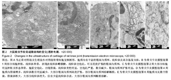
6周后:细胞形态不规则,胞质内见数个脂滴,脂滴体积大,细胞核固缩,大部分线粒体消失,仅有少量线粒体存在,大部分粗面内质网断裂溶解,游离核糖体减少。见图2E。
| [1] Li X, Lang W, Ye H, et al.Tougu Xiaotong capsule inhibits the tidemark replication and cartilage degradation of papain- induced osteoarthritis by the regulation of chondrocyte autophagy. Int J Mol Med. 2013; 31(6):1349-1356. [2] 梁彦勤,廖荣臻,刘超,等.骨性关节炎动物模型研究概况[J].广西中医药大学学报,2012,15(3):64-66. [3] 邓宇,筱梅,任医民,等.关节腔内注射不同蛋白酶建立兔膝骨关节炎模型的对比研究[J].中华关节外科杂志,2009,3(3):332-338. [4] 王庆蓉,官颖鹏,邵卫.软骨细胞在膝骨性关节炎中的超微结构改变[J].电子显微学报,2000,19(6):819-823. [5] 曾庆徐,黄少弼,肖征宇.症状性骨关节炎临床和流行病学探讨[J].中华内科杂志,1995,34(2):88-90. [6] Bendele AM. Animal models of osteoarthritis in an era of molecular biology. JMusculoskelet Neuronal Interact. 2002; 2(6):501-503. [7] Young MF. Mouse models of osteoarhritis provide new research tools. Trends Phamacol Sci.2005;26(7):333-335. [8] 杨峰,史宗道.用木瓜蛋白酶建立兔颞颌关节骨关节炎模型的研究[J].华西口腔医学杂志,2002,20(10):330. [9] Okazaki R,Sakai A,Ootsuyama A,et al.Apoptosis and p53Expresson in Chondrocytes Relate to Degeneration in Articular Cartilage of Immobilized Knee Joints. Rheumatol. 2003;30(3):559-566. [10] Wancket LM,Baragi V,Bove S.Anatomical localization of cartilage degradationmarkers in a surgically induced ratosteoarthritismodel.Toxicologic pathology.2005;33(4):484. [11] Mosikowitiz RW. Experimental models of osteoarthritis. Innosteoarthristis Saunders.Philadelphia.1984;109-115. [12] Bunger C,Hjermind J,Harving S,et al.Relationship between introasseous pressures and intra-articular pressures in arthritis of the knee.Acta Orthop Scand.1983;54(2):188-193. [13] Lozoya KA,Flores JB.A novel rat osteoarthrosis model to assess apoptosis and matrix degradationpathol.Res Pract. 2000;196(11):729-745. [14] 吴宏斌,杜靖远,胡勇,等.兔前交叉韧带切断骨关节炎模型中MMP-1、MMP-13及TIMP-1的mRNA表达研究[J].中华风湿病学杂志,2002,6(3):169. [15] 王健,敖英芳.后交叉韧带断裂继发关节软骨退行性变的实验研究[J].中国运动医学杂志,2004,23(5):476. [16] 顾廷,戴克戎,裘世静,等.应力降低导致关节软骨退变机理的形态学研究[J].中华骨科杂志,1995,15(9):631-633. [17] 毛宾荛.膝关节痛与膝关节骨内压[J].中华骨科杂志,1993, 13(20): 137-139. [18] 陈宝兴,丁继华.双后肢大白鼠的骨关节病实验研究[J].中华骨科杂志,1986,7(2):96. [19] 沈培芝,石印玉.强筋方治疗试验性膝骨关节炎的组织病理学观察研究[J].中国中医骨伤科,1995,3(1):10-13. [20] 路磊,王竞.关节不稳定诱发家兔膝关节骨关节炎的实验研究[J].局解手术学杂志,1995,4(1):23-24. [21] 陈宏贤,王大平,牛琼,等.骨关节炎动物模型的建立及选择[J].深圳中西医结合杂志,2008,18(4):209-212. [22] Aigner T,Cook JL,Gerin N.et al.Histopathology atlas of animal model systems-overview of guiding principles.Osteoarthritis Cartilage.2010;18(3):2-6. [23] Kikuchi T,Sakuta T,Yamaguchi T.Intra-articular injection of collagenase induces experimental osteoarthritis in mature rabbits.Osteoarthritis Cartilage.1998;6(3):177-186. [24] Tsai CL,Liu TK.Estradiol-induced knee osteoarthrosis in ovariectomized rabbits.Clin Orthop.1993;(291):295-302. [25] 邓宇,伍筱梅,任医民,等.关节腔内注射不同蛋白酶建立兔膝骨关节炎模型的对比研究[J].中华关节外科杂志2009,3(3):332-339. [26] Han GY, Ling PX, Wang FS, et al.Comparison study on knee osteoarthritis in rabbits induced by different concentrations of papain,Zhongguo Gu Shang.2012:25(5):424-429. [27] 孙鲁宁,赵燕华,黄桂成.木瓜蛋白酶诱导膝关节骨关节炎模型兔滑膜病理变化与药物注射时间的关系[J].中国组织工程研究与临床康复,2011,15(50):9311-9316. [28] 华英汇,顾湘杰,陈世益,等.威灵仙注射液对骨关节炎影响的实验研究[J].中国运动医学杂志,2003,22(4):420-422. [29] 汪宗保,廖威明,陈朝晖,等.木瓜蛋白酶诱导大鼠膝早期骨关节炎软骨表面的扫描电镜观察[J].中国组织工程研究杂志,2014, 18(2):177-182. [30] Havdrup T,Telhag H.Papain-induced changes in the knee joints of adult rabbits. Acta Orthop Scand.1977,48(2): 143-149. [31] 孙鲁宁,黄桂成,赵燕华,等.木瓜蛋白酶诱导兔膝关节骨关节炎模型滑膜中白细胞介素1、白细胞介素6、白三烯浓度变化与药物注射时间的关系[J].中国组织工程研究,2012,16(33):6184- 6188. [32] 谭庆远,王黎明,曲洪雪,等.中药萃取超导透入治疗兔膝骨关节炎疗效的实验研究[J].中医临床研究,2011,3(3):11-13. [33] 庄超,刘瑞平,徐南伟,等.兔骨关节炎模型血清炎症指标的动态观察[J].南京医科大学学报,2011,31(3):369-373. [34] Vignon E, Arlot M, Vignon G. Etude de al densite cellulaire due cartilage delatete femoral en function delage.Rev Rhum Mal Oeteoarthritic.1976;13(4):365. [35] 娄思权.骨关节炎的病理与发病因素[J].中华骨科杂志,1996, 16(1):56-59. [36] 石辉,何斌,史晨辉,等.用尿激酶型纤溶酶原激活物建立兔骨关节炎模型的研究[J].石河子大学学报,2009,24(1): 66-69. [37] Muehleman C,Green J,Williams JM,et al.The efect of bone remodels inhibition by the oledronnic acid in an animal model of cartilage matrix damage.Osteoarthritis Cartilage.2002; 10(3):226-233. [38] Kikuchi T ,Sakuta T,Yamaguchi T.Intra-articular injection of collagenase induces experimental osteoarthritis in mature rabbits.Osteoarthritis Cartilage.1998;6(3):177-186. [39] 杨峰,史素道.用木瓜蛋白酶建立兔颞颌关节骨关节炎模型[J].华西口腔医学杂志,2002,12(5):330-333. [40] Kopp S, Mejersjo C, Clemenssson E. Induction of osteoarthrosis in the guinea pig knee by papain. Oral Surg Oral Med Oral Pathol. 1983;55(3):259-266. [41] 江捍平,王大平.骨关节炎动物模型[J].中国现代医学杂志2004, 14(6):153-156. [42] 陈百成,张静.骨关节炎[M].北京: 人民卫生出版社,2006: 199-202. [43] 张文贤,张晓刚.骨性关节炎的实验研究进展[J].中国中医骨伤科杂志, 2003,11(6):51-53. [44] 柴本甫,汤雪明.实验性骨关节炎超微结构研究[J].上海第二医科大学学报,1988,8(3):193-198. [45] 唐旭升,杜宁.手法治疗大鼠膝骨关节炎的超微结构研究[J].中医骨伤科杂志,2001,9(2):7-10. [46] 戴七一,文宗振.逍遥散对雌性兔膝关节软骨细胞超微结构的影响[J].中医正骨,2012,24(9):8-10. [47] 刘献祥,李西海,周江涛.改良 Hulth造模法复制膝骨性关节炎的实验研究[J].中国中西医结合杂志,2005,25(12):1104-1108. [48] 张红宇,赵卫东,高岩峰.O3对骨性关节炎关节软骨作用的MR与电镜观察[J].当代医学,2009,15(35):759-761. |
| [1] | 陈子扬, 蒲 锐, 邓 爽, 袁凌燕. 外泌体对运动介导胰岛素抵抗类疾病的调控作用[J]. 中国组织工程研究, 2021, 25(25): 4089-4094. |
| [2] | 陈 扬, 黄邓高, 高元慧, 王顺兰, 曹 卉, 郑琳麟, 何浩伟, 罗思琴, 肖敬川, 张应爱, 张淑芳. 低强度脉冲场超声促进人脂肪间充质干细胞的增殖和黏附[J]. 中国组织工程研究, 2021, 25(25): 3949-3955. |
| [3] | 杨俊辉, 罗金莉, 袁小平. 人生长激素对人牙周膜干细胞增殖及成骨分化的影响[J]. 中国组织工程研究, 2021, 25(25): 3956-3961. |
| [4] | 孙建威, 杨新明, 张 瑛. 孟鲁司特联合骨髓间充质干细胞移植治疗脊髓损伤模型大鼠[J]. 中国组织工程研究, 2021, 25(25): 3962-3969. |
| [5] | 高 珊, 黄东静, 洪海漫, 贾京桥, 孟 斐. 人胎盘间充质干细胞及诱导的胰岛样细胞移植治疗妊娠期糖尿病大鼠效果比较#br#[J]. 中国组织工程研究, 2021, 25(25): 3981-3987. |
| [6] | 郝晓娜, 张英杰, 李玉云, 许 涛. 过表达脯氨酰寡肽酶的骨髓间充质干细胞修复肝纤维化模型大鼠[J]. 中国组织工程研究, 2021, 25(25): 3988-3993. |
| [7] | 刘建友, 贾中伟, 牛佳伟, 曹鑫杰, 张 栋, 魏 杰. 构建股骨3D数字化模型提出一种新的股骨颈前倾角测量方法[J]. 中国组织工程研究, 2021, 25(24): 3779-3783. |
| [8] | 孟令杰, 钱 辉, 盛晓磊, 陆剑锋, 黄建平, 祁连港, 刘宗宝. 3D打印建模联合骨水泥成形微创治疗塌陷Sanders Ⅲ型跟骨骨折[J]. 中国组织工程研究, 2021, 25(24): 3784-3789. |
| [9] | 钱选昆, 黄合飞, 武成聪, 刘克廷, 欧 华, 张金鹏, 任 静, 万建杉. 计算机导航微创经椎间孔腰椎椎间融合治疗腰椎滑脱[J]. 中国组织工程研究, 2021, 25(24): 3790-3795. |
| [10] | 胡 靖, 向 阳, 叶 川, 韩子冀. 3D打印辅助与徒手置钉经皮椎弓根钉内固定治疗胸腰椎骨折的1年随访[J]. 中国组织工程研究, 2021, 25(24): 3804-3809. |
| [11] | 舒启航, 廖亦佳, 薛静波, 晏怡果, 王 程. 新型颈椎3D打印多孔椎间融合器的三维有限元分析[J]. 中国组织工程研究, 2021, 25(24): 3810-3815. |
| [12] | 王一寒, 李 杨, 张 玲, 张 睿, 徐瑞达, 韩晓峰, 程光齐, 王伟力. 数字骨科三维可视化技术在股骨转子间骨折复位内固定中的应用[J]. 中国组织工程研究, 2021, 25(24): 3816-3820. |
| [13] | 孙玛骥, 王秋安, 张星晨, 郭 冲, 袁 峰, 郭开今. 新型颈椎前路经椎弓根固定钉板系统的研制及生物力学分析[J]. 中国组织工程研究, 2021, 25(24): 3821-3825. |
| [14] | 林 旺, 王盈盈, 郭卫中, 袁翠华, 许胜贵, 张申申, 林成寿. 胫骨平台后外侧柱骨折扩大外侧入路内固定增强力学稳定性及膝关节功能[J]. 中国组织工程研究, 2021, 25(24): 3826-3827. |
| [15] | 朱 云, 陈 渝, 邱 皓, 刘 盾, 靳国荣, 陈诗谋, 翁 政. 对侧皮质锁定螺钉治疗骨质疏松股骨骨折的有限元分析[J]. 中国组织工程研究, 2021, 25(24): 3832-3837. |
取材方法:于木瓜蛋白酶首次注射后1,2,4,6周作为取材的时间节点,对SD大鼠行1%氯胺酮腹腔注射麻醉后断头处死,每个时间节点3只。大鼠处死后,膝关节周围2 cm区域剃毛,戴手套、口罩,消毒,铺洞巾,打开膝关节腔,取右膝关节股骨内侧髁关节软骨作为骨关节炎模型组,取左膝关节股骨内侧髁关节软骨作为生理盐水对照组。在第1周时同时切取正常对照组未干预大鼠的右膝关节股骨内侧髁正常软骨组织作为正常对照。见图1。
.jpg)
主要观察指标:大鼠膝关节软骨超微结构的形态学变化。
| 阅读次数 | ||||||
|
全文 |
|
|||||
|
摘要 |
|
|||||