中国组织工程研究 ›› 2021, Vol. 25 ›› Issue (24): 3784-3789.doi: 10.12307/2021.080

• 数字化骨科 digital orthopedics • 上一篇    下一篇

3D打印建模联合骨水泥成形微创治疗塌陷Sanders Ⅲ型跟骨骨折

孟令杰,钱  辉,盛晓磊,陆剑锋,黄建平,祁连港,刘宗宝   

  1. 苏州大学附属张家港医院手足外科,江苏省张家港市   215600
  • 收稿日期:2020-09-24 修回日期:2020-09-28 接受日期:2020-11-11 出版日期:2021-08-28 发布日期:2021-03-05
  • 通讯作者: 刘宗宝,主任医师,硕士生导师,苏州大学附属张家港医院手足外科,江苏省张家港市 215600
  • 作者简介:孟令杰,男,1990年生,2016年苏州大学毕业,硕士,主治医师,主要从事手足显微外科方面的研究。
  • 基金资助:
    江苏大学医学临床科技发展基金(JLY20160120),项目负责人:钱辉

Application of three-dimensional printing technology combined with bone cement in minimally invasive treatment of the collapsed Sanders III type of calcaneal fractures

Meng Lingjie, Qian Hui, Sheng Xiaolei, Lu Jianfeng, Huang Jianping, Qi Liangang, Liu Zongbao   

  1. Department of Hand and Foot Surgery, Zhangjiagang Hospital Affiliated to Soochow University, Zhangjiagang 215600, Jiangsu Province, China
  • Received:2020-09-24 Revised:2020-09-28 Accepted:2020-11-11 Online:2021-08-28 Published:2021-03-05
  • Contact: Liu Zongbao, Chief physician, Master’s supervisor, Department of Hand and Foot Surgery, Zhangjiagang Hospital Affiliated to Soochow University, Zhangjiagang 215600, Jiangsu Province, China
  • About author:Meng Lingjie, Master, Attending physician, Department of Hand and Foot Surgery, Zhangjiagang Hospital Affiliated to Soochow University, Zhangjiagang 215600, Jiangsu Province, China
  • Supported by:
     the Medical Clinical Science and Technology Development Fund of Jiangsu University, No. JLY20160120 (to QH)

摘要:

文题释义:
3D打印(3DP):即快速成型技术的一种,又称增材制造 ,它是一种以数字模型文件为基础,运用粉末状金属或塑料等可黏合材料,通过逐层打印的方式来构造物体的技术。随着技术的发展与成熟,3D打印技术在医学中应用日益增多,从最初的颌面外科及整形外科到目前复杂的骨科手术中,因其个性化的治疗而得到更多重视,已成为满足个性化需求的重要途径。
Sanders Ⅲ型跟骨骨折:Sanders分型是跟骨骨折较为常用的一种分型方式,是基于跟骨冠状面CT扫描,选择跟骨后距关节面最宽处,从外向内将其分为3份(A、B、C),分别代表骨折线位置,这样就有可能有四部分骨折块、三部分关节面骨折块和二部分载距突骨折块。Sanders Ⅲ型是三部分骨折,典型骨折有一中央压缩骨块,即为部分塌陷型跟骨骨折。

背景:跟骨骨折的治疗目前尚存在争议,传统切开复位并发症居高不下,而微创治疗指征较少,应用受限。近年来伴随3D打印技术发展,可否将微创治疗的指征扩大并获得良好效果是跟骨骨折治疗研究的重点与热点。
目的:探讨3D打印技术在骨水泥填充微创治疗塌陷Sanders Ⅲ型跟骨骨折中的应用价值。
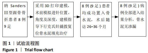
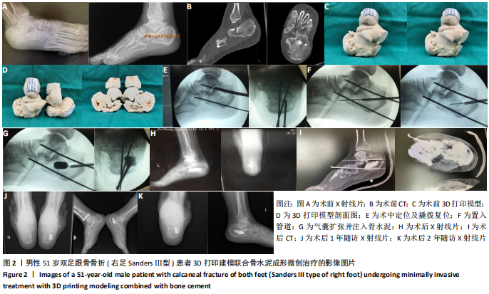
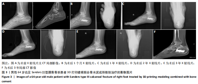
方法:选择苏州大学附属张家港医院手足外科2016年9月至2017年4月收治的8例9足Sanders Ⅲ型跟骨骨折患者,全部为男性,年龄50-66岁,平均57.4岁。所有患者均进行患肢CT检查,并获得3D打印个体化后足部模型,模拟撬拨进针位置及及进针角度,在撬拨复位辅助下行骨水泥填充。记录所有患者术前、术后的Bohler角、Gissane角以及跟骨长度、宽度、高度;术后功能评价参照美国足踝外科协会后足评分系统。
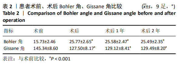
结果与结论:①所有患者均得到随访,随访时间26-36个月,骨折全部愈合,无再次塌陷发生,骨水泥无渗漏,未出现排异反应,无皮肤坏死及感染,跟骨外形恢复满意,足部功能良好;②美国足踝外科协会后足评分优7例,良1例,差1例,优良率为89%;③患者Bohler角、Gissane角术前为(15.73±2.46)°和(145.34±8.6)°,术后改善至(25.77±2.65)°和(127.5±8.17)°,差异有显著性意义(P < 0.001);术后Bohler角、Gissane角与术后1,2年对比差异均无显著性意义(P > 0.05);④提示3D打印个体化建模联合骨水泥填充治疗塌陷的Sanders Ⅲ型跟骨骨折效果良好,具有个体化、针对性强、手术损伤小、并发症少等优点。
https://orcid.org/0000-0001-6944-9621 (孟令杰) 

中国组织工程研究杂志出版内容重点:人工关节;骨植入物;脊柱;骨折;内固定;数字化骨科;组织工程

关键词: 跟骨骨折, 3D打印, 骨水泥, 撬拨复位, 微创

Abstract: BACKGROUND: The treatment of calcaneal fracture is still controversial at present. Traditional complications of open reduction remain high, while minimally invasive treatment has few indications and its application is limited. In recent years, with the development of three-dimensional (3D) printing technology, whether the indications of minimally invasive treatment can be expanded and achieve good results or not is the focus and hot spot of study on the treatment of calcaneal fracture.  
OBJECTIVE: To explore the application value of 3D printing technology in the minimally invasive treatment of collapsed Sanders III calcaneal fractures.
METHODS:  Eight patients with nine foot Sanders III calcaneal fractures who were admitted to the Department of Hand and Foot Surgery of Zhangjiagang Hospital Affiliated to Soochow University from September 2016 to April 2017. All were males, aged 50-66 years, with an average age of 57.4 years. All patients underwent CT examination and obtained 3D printed personalized posterior foot, followed by bone cement filling surgery assisted by pry reduction after simulating the position and angle of pry needle. The length, width and height of calcaneu and the Gissane angle and Bohler angle before and after surgery of all patients were recorded. The postoperative function was evaluated according to the American Orthopedic Foot Ankle Society Ankle Hindfoot Scale.  
RESULTS AND CONCLUSION: (1) All patients were followed up for 26-36 months. All fractures healed. There was no collapse, leakage of bone cement, rejection reaction, skin necrosis or infection. Calcaneal shape recovered satisfactorily, and foot function was good. (2) American Orthopedic Foot Ankle Society Ankle Hindfoot Scale results showed excellent in seven cases, good in one case, and poor in one case, with excellent and good rate of 89%. (3) Bohler angle and Gissane angle of patients respectively improved from (15.73±2.46)° and (145.34±8.6)° before surgery to (25.77±2.65)° and (127.5±8.17)° after surgery. Difference was statistically significant (P < 0.001). There was no significant difference in Bohler angle and Gissane angle between immediately, and 1, 2 years after operation (P > 0.05). (4) It is indicated that 3D printing individualized bone cement filling plays an important clinical role in the treatment of collapsed Sanders III calcaneal fracture and it has the advantages of short time of individualized targeted operation, minor surgical injury, and few complications.

Key words: calcaneal fracture, 3D printing, bone cement, pry reduction, minimally invasive

中图分类号: