| [1] Galibert P, Deramond H, Rosat P, et al. Preliminary note on the treatment of vertebral angioma by percutaneous acrylic vertebroplasty.Neurochirurgie. 1987;33:166-167.
[2] 刘小勇,杨惠林. Kyphoplasty经皮定位技术的解剖研究与临床应用[J].苏州大学学报,2004, 1(15):40-45.
[3] 马建华,王庆雷,杨兆义,等.改良椎体成形术的临床探讨[J].生物骨科材料与临床研究,2011,8(2):15.
[4] 刘尚礼,叶伟,李春海.经皮椎体成形术的研究进展[J].脊柱外科杂志,2008,6(1):58-61.
[5] 赵俊强,陈琼.单侧与双侧经皮椎体成形术治疗椎体压缩性骨折的前瞻性研究[J].中国骨与关节损伤杂志,2011,26(3):229-230.
[6] Garfin SR, Yuan HA, Reiley MA. New technologies in spine:KyhoPlasty and Vertebroplasty for the treatment of painful osteopotic compression fractures.Spine. 2001; 26(14): 1511-1515.
[7] 刘小勇,杨惠林,唐天驷,等.椎体后凸成形术棘突定位穿刺点与穿刺轨道的研究[J].中华骨科杂志,2005,25(8): 462-463.
[8] 孟斌,杨惠林,黄晨,等.手术技术对椎体后凸成形术疗效的影响[J].山东医药,2010,50(46): 71.
[9] 吴红言. 经皮穿刺椎体成形术的手术配合体会[J]. 临床护理, 2011,49(3):52-53.
[10] 杨升全,金正帅,张宁.椎体扩张器、Kyphon球囊和Sky骨膨胀器三种后凸成形术的临床应用比较研究[J].南京医科大学学报, 2011,31(2):251-252.
[11] Ebraheim NA,Xu R, Ahmad M, et al. Projeetion of the thoracie Pedicle and its Morphometrie analysis. Spine. 1997;22(3): 233-238.
[12] 赵士军,高景春,宓士军,等.单侧入路胸椎椎体成形术治疗胸椎骨折[J].临床医药实践,2011,20(6):44-45.
[13] 戴尅戎,荣国威.骨折治疗的AO原则[M].北京:人民卫生出版社, 2005:262-263.
[14] 李龙,李兵,苟凌云,等.经皮椎体成形术治疗胸腰椎骨质疏松性压缩性骨折[J].中华微创外科杂志,2007,7(7):621-622.
[15] 滕皋军,何仕诚,郭金和.经皮椎体成形术治疗椎体良恶性病变的临床技术应用探讨[J].中华放射学杂志,2002,36(4):295-299.
[16] 李子祥,李绍科,刘丰春.与经皮椎体成形术相关的L5椎弓根影像解剖学研究[J].临床解剖学杂志,2007,25(2):160-162.
[17] 徐志强,邓忠良,胡永军,等.椎弓根基底部穿刺角度的测量和临床意义[J].重庆医科大学学报,2007,32(7):735-738.
[18] 赵俊强, 陈琼.单侧与双侧经皮椎体成形术治疗椎体压缩性骨折的前瞻性研究[J].骨与关节损伤杂志,2011,26(3):229-230.
[19] Steffee AD, Biscup RS, Stkowski DJ. Segmental Spine Plates with Pedicle screw fixation a new intemal fixation device for disorders of the lumbar and thoracolumbar. Spine Clin Orthop. 1986;203:45-53.
[20] 杜心如,叶启彬.经推弓根胸腰推内固定应用解剖学研究的进展[J].中国矫形外科杂志,1998,5(5):446-448.
[21] Phillips JH, Kling TF Jr, Cohen MD. The radiographic anatomy of the thoracic pedicle. Spine (Phila Pa 1976). 1994;19(4):446-449.
[22] Weinstein JN, Spratt KF, Spengler D, et al. Spinal pedicle fixation: reliability and validity of roentgenogram-based assessment and surgical factors on successful screw placement. Spine (Phila Pa 1976). 1988;13(9):1012-1018.
[23] 石锐,袁元.不同节段椎弓根内部结构的测量和比较[J].中国临床解剖学杂志,2005,23(5):458-462.
[24] Miehele AA, Krueger FJ. Surgical approach to the vertebral body. J Bone Joint Surg Am. 1949;31A(4):873-878.
[25] Krag MH, Beynnon BD, Pope MH, et al. An internal fixator for posterior application to short segments of the thoracic, lumbar, or lumbosacral spine. Design and testing. Clin Orthop Relat Res. 1986;(203):75-98. |
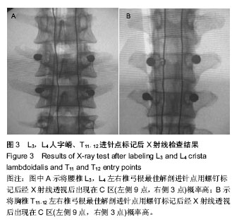

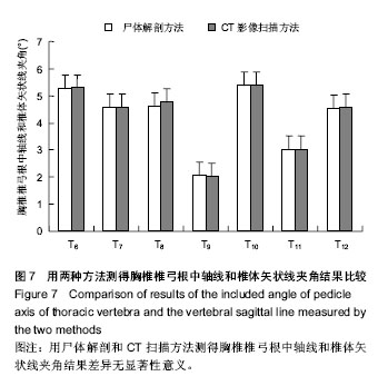
.jpg)
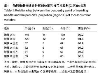
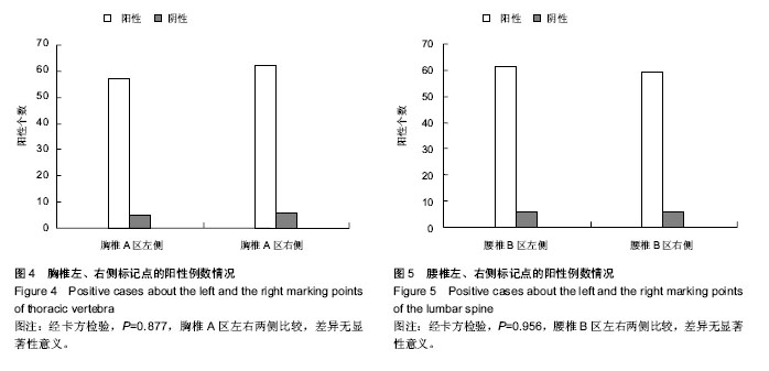
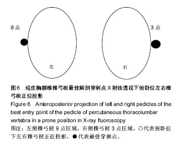
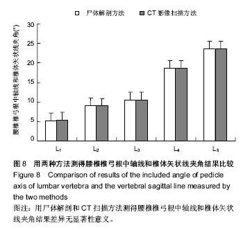
.jpg)