| [1] Salzar RS, “Dale” Bass CR, Kent R, et al. Development of injury criteria for pelvic fracture in frontal crashes. Traffic Inj Prev. 2006;7(3): 299-305.
[2] Hammel J, Legome E. Pelvic fracture. J Emerg Med. 2006; 30(1): 87-92.
[3] Ponsen KJ, Joosse P, Schigt A, et al. Internal fracture fixation using the Stoppa approach in pelvic ring and acetabular fractures: technical aspects and operative results. J Trauma Acute Care Surg. 2006;61(3): 662-667.
[4] Suzuki T, Shindo M, Soma K, et al. Long-term functional outcome after unstable pelvic ring fracture. J Trauma Acute Care Surg. 2007;63(4): 884-888.
[5] Citak M, Gardner MJ, Kendoff D, et al. Virtual 3D planning of acetabular fracture reduction. J Orthop Res. 2008;26(4): 547-552.
[6] Khoury A, Kreder H, Skrinskas T, et al. Lateral compression fracture of the pelvis represents a heterogeneous group of complex 3D patterns of displacement. Injury. 2008;39(8): 893-902.
[7] Øvre S, Madsen JE, Røise O. Acetabular fracture displacement, roof arc angles and 2 years outcome. Injury. 2008;39(8): 922-931.
[8] Davis JW, Moore FA, McIntyre Jr RC, et al. Western trauma association critical decisions in trauma: management of pelvic fracture with hemodynamic instability. J Trauma Acute Care Surg. 2008;65(5): 1012-1015.
[9] Lefaivre KA, Padalecki JR, Starr AJ. What constitutes a Young and Burgess lateral compression-I (OTA 61-B2) pelvic ring disruption? A description of computed tomography-based fracture anatomy and associated injuries. J Orthop Trauma. 2009;23(1):16-21.
[10] Carmack DB, Moed BR, McCarroll K, et al. Accuracy of detecting screw penetration of the acetabulum with intraoperative fluoroscopy and computed tomography. J Bone Joint Surg. 2001;83(9): 1370-1375.
[11] 高巍,彭阿钦,陈百成,等.髋臼螺钉固定安全性的解剖学研究[J]. 中国矫形外科杂志,2005, 13(11): 840-842.
[12] 王先泉,张进禄,周东生.髋臼后柱支持钢板的临床解剖学研究[J]. 中国骨与关节损伤杂志, 2005, 20(1): 9-11.
[13] 张本,苏佳灿,王瑞官,等.髋臼后柱解剖学测量及其临床意义[J]. 国际骨科学杂志,2006, 27(3):186-188.
[14] 牛云飞,王家林,张春才.髋臼前壁厚度的解剖学测量及其意义[J].中国骨与关节损伤杂志,2007, 22(6): 459-461.
[15] 牛云飞,许硕贵,张春才.髋臼后壁厚度的解剖学测量及其意义[J]. 中国临床解剖学杂志, 2007, 25(4): 400-402.
[16] Chiu FY, Chen CM, Lo WH. Surgical treatment of displaced acetabular fractures-72 cases followed for 10 (6–14) years. Injury. 2000;31(3): 181-185.
[17] 张奉琪,潘进社,张英泽,等.骨盆骨折血管损伤的解剖学基础[J]. 中国临床解剖学杂志,2004, 22(2): 116-119.
[18] 张鹏,王家林,禹宝庆,等.影响髋臼骨折手术预后的因素分析[J]. 国际骨科学杂志, 2006, 27(2): 75-77.
[19] Haidukewych GJ, Scaduto J, Herscovici Jr D, et al. Iatrogenic nerve injury in acetabular fracture surgery: a comparison of monitored and unmonitored procedures. J Orthop Trauma. 2002;16(5): 297-301.
[20] Gruson KI, Moed BR. Injury of the femoral nerve associated with acetabular fracture. J Bone Joint Surg. 2003; 85(3): 428-431.
[21] 阮默,徐达传,汪新民,等.经皮骶髂螺钉内固定术的应用解剖学研究[J].中国临床解剖学杂志,2006, 24(5): 479-484.
[22] 宋磊,李严兵,王平山,等.经皮手术治疗骶髂关节损伤的临床解剖学数字化研究[J].中国矫形外科杂志, 2007, 15(10): 765-767.
[23] 蓝旭,梁军,文益民,等.髋臼骨折手术并发症分析[J].中国现代手术学杂志, 2007, 11(5): 373-376.
[24] 张浩,沙强,田禾,等.髋臼骨折手术并发症及防治[J].中国现代医学杂志, 2007, 17(24): 3064-3067.
[25] Ebraheim NA, Xu R, Biyani A, et al. Anatomic basis of lag screw placement in the anterior column of the acetabulum. Clin Orthop Related Res. 1997;339: 200-205.
[26] 王庆贤,张英泽,潘进社,等.髋臼前柱拉力螺钉内固定的定量解剖学研究[J].中国临床解剖学杂志, 2004, 22(2): 133-135.
[27] Anglen JO, DiPasquale T. The reliability of detecting screw penetration of the acetabulum by intraoperative auscultation. J Orthop Trauma. 1994;8(5): 404-408.
[28] Ebraheim NA, Savolaine ER, Hoeflinger MJ, et al. Radiological diagnosis of screw penetration of the hip joint in acetabular fracture reconstruction. J Orthop Trauma. 1989; 3(3): 196-201.
[29] Carmack DB, Moed BR, McCarroll K, et al. Accuracy of detecting screw penetration of the acetabulum with intraoperative fluoroscopy and computed tomography. J Bone Joint Surg. 2001;83(9): 1370-1375.
[30] Shazar N, Brumback RJ, Novak VP, et al. Biomechanical evaluation of transverse acetabular fracture fixation. Clin Orthop Related Res. 1998;352: 215-222.
[31] Shiramizu K, Naito M, Yatsunami M. Quantitative anatomic characterisation of the pelvic brim to facilitate internal fixation through an anterior approach. J Orthop Surg (Hong Kong). 2003;11(2):137-140.
[32] Benedetti JA, Ebraheim NA, Xu R, et al. Anatomic considerations of plate-screw fixation of the anterior column of the acetabulum. J Orthop Trauma. 1996;10(4): 264-272.
[33] 王先泉,张伟,孙水,等.髋臼前柱骨折钢板中内固定的最佳进针点[J].中国组织工程研究与临床康复, 2009 ,13(17): 3232-3236. |
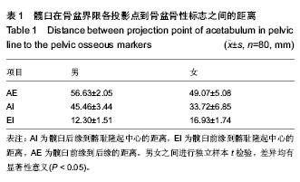


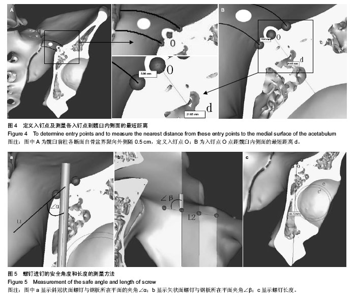
.jpg)
.jpg)
.jpg)