中国组织工程研究 ›› 2014, Vol. 18 ›› Issue (18): 2830-2835.doi: 10.3969/j.issn.2095-4344.2014.18.008
• 器官移植动物模型 organ transplantation and animal model • 上一篇 下一篇
兔适合构建鼻窦炎动物模型
解道宇1,鞠建宝1,于海玲1,李 娜1,郝大鹏2,隋爱华3
- 青岛大学医学院附属医院,1耳鼻咽喉头颈外科,2放射科,3中心实验室,山东省青岛市 266003
-
收稿日期:2014-02-13出版日期:2014-04-30发布日期:2014-04-30 -
通讯作者:于海玲,医学博士,副教授,硕士生导师,青岛大学医学院附属医院耳鼻咽喉头颈外科,山东省青岛市 266003 -
作者简介:解道宇,男,1984年生,山东省菏泽市人,汉族,青岛大学医学院在读硕士,主要从事鼻科学及鼻内镜技术的基础与临床研究工作。 -
基金资助:山东省高等学校科技计划项目(J13LL56)
Rabbit is suitable for establishing an animal model of rhino-sinusitis
Xie Dao-yu1, Ju Jian-bao1, Yu Hai-ling1, Li Na1, Hao Da-peng2, Sui Ai-hua3
- 1Department of Otolaryngology-Head and Neck Surgery, Affiliated Hospital of Qingdao University Medical College, Qingdao 266003, Shandong Province, China
2Medical Imaging, Affiliated Hospital of Qingdao University Medical College, Qingdao 266003, Shandong Province, China
3Central Laboratory, Affiliated Hospital of Qingdao University Medical College, Qingdao 266003, Shandong Province, China
-
Received:2014-02-13Online:2014-04-30Published:2014-04-30 -
Contact:Yu Hai-ling, M.D., Associate professor, Master’s supervisor, Department of Otolaryngology-Head and Neck Surgery, Affiliated Hospital of Qingdao University Medical College, Qingdao 266003, Shandong Province, China -
About author:Xie Dao-yu, Studying for master’s degree, Department of Otolaryngology-Head and Neck Surgery, Affiliated Hospital of Qingdao University Medical College, Qingdao 266003, Shandong Province, China -
Supported by:the Science and Technology Project of Higher Learning Institutes of Shandong Province, No. J13LL56
摘要:
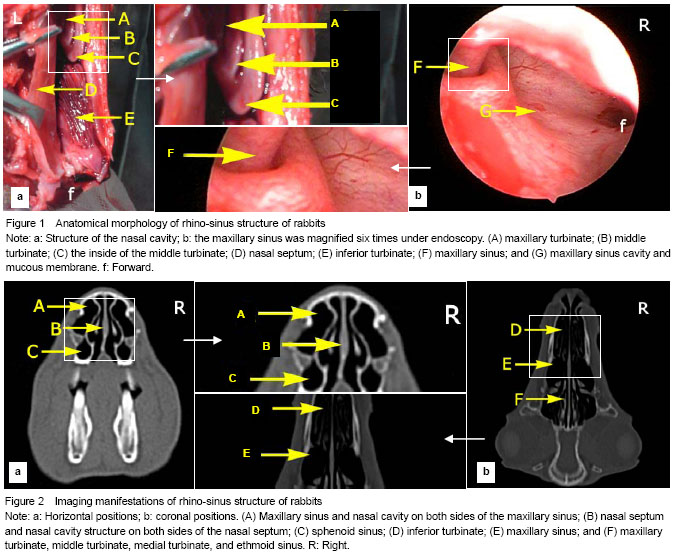
背景:以实验动物模拟人类相关疾病是研究疾病发生发生及治疗的基础,鼻腔、鼻窦疾病也需要恰当实验动物作为其模型。 目的:观察兔鼻腔、鼻窦的CT和局部解剖表现,探讨兔应用于鼻窦炎动物模型的可行性。 方法:利用CT使用常规鼻窦冠状位与水平位扫描新西兰兔,随后进行常规鼻腔、鼻窦解剖学观察。 结果与结论:兔鼻中隔将鼻腔分为左右两个腔,鼻腔外侧壁由上颌鼻甲、中间鼻甲、内侧鼻甲、下鼻甲组成,上颌窦窦腔最大,筛窦、蝶窦、额窦相对较小,以上结构均对称分布。兔鼻腔、鼻窦在CT扫描下显示清晰。兔鼻腔、鼻窦的解剖与人类鼻腔、鼻窦解剖结构既有相似也有不同,其上颌窦解剖部位与人相似且窦腔较大便于操作,具有动物实验模型建立的可行性,适用于鼻窦炎动物模型建立,可应用于模拟人类鼻窦炎的研究。
中图分类号:
引用本文
解道宇,鞠建宝,于海玲,李 娜,郝大鹏,隋爱华. 兔适合构建鼻窦炎动物模型[J]. 中国组织工程研究, 2014, 18(18): 2830-2835.
Xie Dao-yu, Ju Jian-bao, Yu Hai-ling, Li Na, Hao Da-peng, Sui Ai-hua. Rabbit is suitable for establishing an animal model of rhino-sinusitis[J]. Chinese Journal of Tissue Engineering Research, 2014, 18(18): 2830-2835.
Quantitative analysis of experimental animals
The bilateral nasal cavity, sinus and nasal bone are shown clearly in the CT scans. The nasal septum is located on both sides of the nasal cavity as the human beings. The turbinate structure is visible at the lateral nasal, and a low-density mass shadow can be observed at both sides in front of lateral wall of the nasal cavity. The anatomical area of the maxillary sinus is shallow, and the sinus cavity with longitudinal elliptic shape is the largest. The ethmoid sinus is in the posterior high side of the maxillary sinus and consists of many air cells. Sphenoid and frontal sinuses are small (Figure 2).
| [1] Kerschner JE, Cruz MJ, Beste DJ, et al. Computed tomography vs. magnetic resonance imaging of acute bacterial sinusitis: a rabbit model. Am J Otolaryngol. 2000; 21(5):298-305. [2] Okuyemi KS, Tsue TT. Radiologic imaging in the management of sinusitis. Am Fam Physician. 2002;66(10): 1882-1886. [3] Aribandi M, Bazan C 3rd. CT and MRI features in Bipolaris fungal sinusitis. Australas Radiol. 2007;51(2):127-132. [4] Diament MJ. The diagnosis of sinusitis in infants and children: x-ray, computed tomography, and magnetic resonance imaging. Diagnostic imaging of pediatric sinusitis. J Allergy Clin Immunol. 1992;90(3 Pt 2):442-444. [5] Mafee MF, Tran BH, Chapa AR. Imaging of rhinosinusitis and its complications: plain film, CT, and MRI. Clin Rev Allergy Immunol. 2006;30(3):165-186. [6] Ide C, Trigaux JP, Eloy P. Chronic sinusitis: the role of imaging. Acta Otorhinolaryngol Belg. 1997;51(4):247-258. [7] Dessi P, Champsaur P, Paris J, et al. Imaging of the adult sinusitis: indications for using conventional techniques, CT scan and MRI. Rev Laryngol Otol Rhinol (Bord). 1999; 120(3):173-176. [8] Goldstein JH1, Phillips CD. Current indications and techniques in evaluating inflammatory disease and neoplasia of the sinonasal cavities. Curr Probl Diagn Radiol. 1998;27(2):41-71. [9] Müller NL. Computed tomography and magnetic resonance imaging: past, present and future. Eur Respir J Suppl. 2002; 35:3s-12s. [10] Schmidt GP, Kramer H, Reiser MF, et al. Whole-body magnetic resonance imaging and positron emission tomography-computed tomography in oncology. Top Magn Reson Imaging. 2007;18(3):193-202. [11] Moulin G, Pascal T, Jacquier A, et al. Radiologic imaging of chronic sinusitis in the adult. J Radiol. 2003;84(7-8 Pt 2): 901-919. [12] Bipat S, Glas AS, van der Velden J, et al. Computed tomography and magnetic resonance imaging in staging of uterine cervical carcinoma: a systematic review. Gynecol Oncol. 2003;91(1):59-66. [13] Schwickert HC, Cagil H, Kauczor HU, et al. CT and MRT of the paranasal sinuses. Aktuelle Radiol. 1994;4(2):88-96. [14] Bertrand B, Collet S, Betsch C, et al. Diagnostic techniques in chronic sinusitis: endoscopy, sinusomanometry. Acta Otorhinolaryngol Belg. 1997;51(4):259-269. [15] Druce HM. The use of ultrasound as an imaging technique in the diagnosis of sinusitis. N Engl Reg Allergy Proc. 1988; 9(2):109-112. [16] Drettner B, Johansson P, Kumlien J. Experimental acute sinusitis in rabbit. A study of mucosal blood flow. Acta Otolaryngol. 1987;103(5-6):432-434. [17] Johansson P, Kumlien J. Blood flow in the rabbit maxillary sinus mucosa during experimentally induced acute sinusitis. Acta Otolaryngol. 1988;106(3-4):299-305. [18] Knowlton CD, McGregor GW. How and when the mucous membrane of the maxillary sinus regenerates: an experimental study in the dog. Arch Otolaryngol. 1928; 8(6):647-656. [19] Hiding A. Experimental sinus surgery: effect of operative windows on normal sinuses. Ann Otol Rhinol Laryngol. 1941(50):379-392. [20] Bomer K, Brichta A, Baroody F, et al. A mouse model of acute bacterial rhinosinusitis. Arch Otolaryngol Head Neck Surg. 1998;124(11):1227-1232. [21] Wei H, Zhu Z, Liu Z, et al. Observation on ultrastructure of maxillary sinus mucosa in experimental acute sinusitis. Lin Chung Er Bi Yan Hou Tou Jing Wai Ke Za Zhi. 2012; 26(11): 519-522. [22] Lin B, Wang HG. Effects of different treating ways to the ostiomeatal complex on changes in ciliary morphology and function of sinuous mucosa in rabbits with experimental chronic sinusitis. Zhongguo Zhongxiyi Jiehe Er Bi Yanhou Ke Zazhi. 2011;19(4):226-230. [23] Jin MZ. Mouse model of acute bacterial rhinosinusitis. Shenyang: China Medical University, 2010. [24] Yu CJ, Lu L, Gu YJ, et al. Development of a rat model of chronic rhinosinusitis and observation of biological characteristics. Zhongguo ErBi Yanhou Ludi Waike Zazhi. 2011;17(6):419-424. [25] Lin B, Wang HG, Lin XQ, et al. Comparison and improvement in the methods of establishing animals model of experimental chronic sinusitis in rabbits. Zhonghua Er Bi Yan Hou Tou Jing Wai Ke Za Zhi. 2005; 40(5):343-346. [26] Kara CO, Demirkan N. A review on experimental sinusitis models in rabbits. Kulak Burun Bogaz Ihtis Derg. 2003; 10(3):122-130. [27] Kara CO. Animal models of sinusitis: relevance to human disease. Curr Allergy Asthma Rep. 2004;4(6):496-499. [28] Naclerio R, Blair C, Yu X, et al. Allergic rhinitis augments the response to a bacterial sinus infection in mice: A review of an animal model. Am J Rhinol. 2006;20(5):524-533. [29] Norlander T, Westrin KM, Stierna P. The inflammatory response of the sinus and nasal mucosa during sinusitis: implications for research and therapy. Acta Otolaryngol Suppl. 1994;515:38-44. [30] Sandler NA, Johns FR, Braun TW. Advances in the management of acute and chronic sinusitis. J Oral Maxillofac Surg. 1996;54(8):1005-1013. [31] Forsgren K, Westrin KM, Fukami M, et al. Effects of surgery on mucosal pathologic changes following experimental sinusitis in rabbit. Ann Otol Rhinol Laryngol. 1998;107(2): 155-163. [32] Hinni ML, McCaffrey TV, Kasperbauer JL. Early mucosal changes in experimental sinusitis. Otolaryngol Head Neck Surg. 1992;107(4):537-548. [33] Liang KL, Jiang RS, Wang J, et al. Developing a rabbit model of rhinogenic chronic rhinosinusitis. Laryngoscope. 2008;118(6):1076-1081. [34] Bolger WE, Leonard D, Dick EJ Jr, et al. Gram negative sinusitis: a bacteriologic and histologic study in rabbits. Am J Rhinol. 1997;11(1):15-25. [35] Zhang J, Zhou B, Zhang SZ, et al. Histopathological observation of bone remodeling in rabbit sinusitis model. Zhonghua Er Bi Yan Hou Tou Jing Wai Ke Za Zhi. 2009; 44(7):546-550. [36] Wang J, Liu F, Ge QJ. A new method to create experimental model of noninvasive fungal sinusitis in rabbits. Dier Junyi Daxue Xuebao. 2012;33(1):16-20. [37] Wei HQ, Li ZQ, Wang QP, et al. Comparison of two methods for establishing a model of experimental acute sinusitis in rabbits. Zhongguo Bijiao Yixue Zazhi. 2007; 17(4):201-205. [38] Casteleyn C, Cornillie P, Hermens A, et al. Topography of the rabbit paranasal sinuses as a prerequisite to model human sinusitis. Rhinology. 2010;48(3):300-304. [39] Kara CO, Cetin CB, Demirkan N, et al. Experimental sinusitis in a rhinogenic model. Laryngoscope. 2004;114(2): 273-278. [40] Khalid AN, Hunt J, Perloff JR, et al. The role of bone in chronic rhinosinusitis. Laryngoscope. 2002;112(11): 1951-1957. [41] Irvin CG. Sinusitis and asthma: an animal model. J Allergy Clin Immunol. 1992;90(3 Pt 2):521-533. [42] Van Bruaene N, Bachert C. Tissue remodeling in chronic rhinosinusitis. Curr Opin Allergy Clin Immunol. 2011;11(1): 8-11. [43] Babbel RW, Harnsberger HR. A contemporary look at the imaging issues of sinusitis: sinonasal anatomy, physiology, and computed tomography techniques. Semin Ultrasound CT MR. 1991;12(6):526-540. [44] Roithmann R, Shankar L, Hawke M, et al. CT imaging in the diagnosis and treatment of sinus disease: a partnership between the radiologist and the otolaryngologist. J Otolaryngol. 1993;22(4):253-260. [45] Dong Y, Zhou B, Wang CS, et al. CT and MRI diagnosis of lesions in unilateral maxillary sinus. Zhonghua Er Bi Yanhou Toujing Waike Zazhi. 2011;18(3):320-325. [46] Ozcan KM, Ozcan I, Selcuk A, et al. Comparison of Histopathological and CT Findings in Experimental Rabbit Sinusitis. Indian J Otolaryngol Head Neck Surg. 2011;63(1): 56-59. [47] Kelemen G. The nasal and paranasal cavities of the rabbit in experimental work. AMA Arch Otolaryngol. 1955;61(5): 497-512. [48] Ding GQ, Zheng CQ. Experimental animal model of sinusitis. Guoji Er Bi Yanhou Tou Jing Wai Ke Zazhi. 2006; 30(4):255-258. [49] Casteleyn C, Cornillie P, Hermens A, et al. Topography of the rabbit paranasal sinuses as a prerequisite to model human sinusitis. Rhinology. 2010;48(3):300-304. [50] Marks SC. Acute sinusitis in the rabbit model: histologic analysis. Laryngoscope. 1998;108(3):320-325. [51] Islam MN, Kundu PK, Biswas MC, et al. Bacteriological profile in chronic dacryocystitis. J Indian Med Assoc. 2006; 104(7):398, 400. [52] Altan-Yaycioglu R, Canan H, Sizmaz S, et al. Nasolacrimal duct obstruction: clinicopathologic analysis of 205 cases. Orbit. 2010;29(5):254-258. [53] Frame NJ, Burkat CN. Identifying an appropriate animal model for the nasolacrimal drainage system. Ophthal Plast Reconstr Surg. 2009;25(5):354-358. [54] Florin M, Rusanen E, Haessig M, et al. Clinical presentation, treatment, and outcome of dacryocystitis in rabbits: a retrospective study of 28 cases (2003-2007). Vet Ophthalmol. 2009;12(6):350-356. [55] Lv P, Li ST, Mang LL, et al. Effect of acupuncture plus oxygen therapy on serum IL-9 and TNF-α in chronic rhinosinusitis rabbits. Shanghai Zhenjiu Zazhi. 2011;30(8): 574-576. [56] Dufour X, Kauffmann-Lacroix C, Goujon JM, et al. Experimental model of fungal sinusitis: a pilot study in rabbits. Ann Otol Rhinol Laryngol. 2005;114(2):167-172. [57] Chakrabarti A, Jatana M, Sharma SC. Rabbit as an animal model of paranasal sinus mycoses. J Med Vet Mycol. 1997; 35(4):295-297. [58] Ge Y, Tsukatani T, Nishimura T, et al. Cell death of olfactory receptor neurons in a rat with nasosinusitis infected artificially with Staphylococcus. Chem Senses. 2002;27(6): 521-527. [59] Bleier BS, Kofonow JM, Hashmi N, et al. Antibiotic eluting chitosan glyceraophosphate implant in the setting of acute bacterial sinusitis: a rabbit model. Am J Rhinol Allergy. 2010; 24(2):129-132. [60] Costa HO, Ruschi e Luchi GE, Augusto AG, et al. Comparative study of several sinusitis experimental modelling techniques in rabbits. Braz J Otorhinolaryngol. 2007;73(5):627-631. |
| [1] | 王德斌, 毕郑刚. 尺骨鹰嘴骨折-脱位解剖力学、损伤特点、固定修复及3D技术应用的相关问题[J]. 中国组织工程研究, 2021, 25(9): 1446-1451. |
| [2] | 吴 训, 孟娟红, 张建运, 王 亮. 浓缩生长因子修复兔髁突全层软骨损伤[J]. 中国组织工程研究, 2021, 25(8): 1166-1171. |
| [3] | 樊佳兵, 张军梅. 成年女性不同垂直骨面型下颌骨形态的测量分析[J]. 中国组织工程研究, 2021, 25(8): 1177-1183. |
| [4] | 唐 辉, 姚志浩, 罗道文, 彭双麟, 杨双林, 王 浪, 肖金刚. 高脂高糖饮食结合链脲佐菌素建立2型糖尿病性骨质疏松症大鼠模型[J]. 中国组织工程研究, 2021, 25(8): 1207-1211. |
| [5] | 陈继铭, 吴晓静, 刘田丰, 陈海聪, 黄成硕. 水飞蓟素对四氯化碳致小鼠肝损伤和骨代谢的影响[J]. 中国组织工程研究, 2021, 25(8): 1224-1228. |
| [6] | 袁新平, 邵艳波, 吴 超, 汪剑龄, 童梁成, 李 颖. 骨折端CT扫描参数个性化微分建模仿真目标骨段的准确性[J]. 中国组织工程研究, 2021, 25(6): 912-916. |
| [7] | 张 经, 王 斌, 吕 欣. 解剖型髓内钉在四肢管状骨骨折治疗中的应用:把持与抗旋转能力更强[J]. 中国组织工程研究, 2021, 25(6): 917-922. |
| [8] | 李 琨 , 李志军 , 张少杰 , 高 尚 , 孙 昊 , 杨 喜 , 王 星 , 戴丽娜. 有限元动态仿真建立4岁儿童“枕-寰-枢”关节模型[J]. 中国组织工程研究, 2021, 25(24): 3773-3778. |
| [9] | 田 洋, 唐 超, 廖烨晖, 唐 强, 马 飞, 钟德君. CT和MRI在腰椎管狭窄症中测量椎管面积的一致性和可重复性[J]. 中国组织工程研究, 2021, 25(24): 3882-3887. |
| [10] | 易美芝, 罗光华, 肖雅雯, 胡 蓉, 陈小龙, 赵 衡. 距骨解剖变异的MRI表现[J]. 中国组织工程研究, 2021, 25(24): 3888-3893. |
| [11] | 周元博, 王晋东. 股骨滑车发育不良的病因及治疗:是先天基因还是后天髌骨应力刺激[J]. 中国组织工程研究, 2021, 25(24): 3908-3913. |
| [12] | 杨 威, 陈泽华, 易志勇, 黄旭东, 韩清民, 张荣华. 关节腔内注射透明质酸与安慰剂治疗早中期膝骨关节炎的疗效差异:基于随机、双盲、对照、临床试验的Meta分析[J]. 中国组织工程研究, 2021, 25(23): 3760-3766. |
| [13] | 解景舒, 张祥林, 刘金磊, 文 静. High Resolution精密断层重建在中、内耳CT扫描成像中的应用[J]. 中国组织工程研究, 2021, 25(23): 3614-3618. |
| [14] | 高 坤, 陈大宇, 张 勇, 刘伟东, 孙淑芬, 赖文强, 马笃军, 吴益宏, 林展鹏, 蒋鹰鹭, 余伟吉. 牛膝醇提物调控滑膜成纤维细胞外泌体抑制软骨细胞外基质降解[J]. 中国组织工程研究, 2021, 25(23): 3636-3640. |
| [15] | 林天烨, 杨 鹏, 熊冰朗, 何晓铭, 颜新昊, 张 进, 何 伟, 魏秋实. 术中画线法与三维重建术前模拟测量股骨截骨旋转角度对比的体外实验[J]. 中国组织工程研究, 2021, 25(21): 3349-3353. |
Design
Time and setting
|
||||||||||||
Rhino-sinus morphology in rabbits was observed.
| 阅读次数 | ||||||
|
全文 |
|
|||||
|
摘要 |
|
|||||