中国组织工程研究 ›› 2013, Vol. 17 ›› Issue (40): 7028-7033.doi: 10.3969/j.issn.2095-4344.2013.40.002

• 骨髓干细胞 bone marrow stem cells • 上一篇    下一篇

Ca 2+参与骨髓间充质干细胞存活增殖及向肝细胞的诱导分化

焦淑贤,胡  彬,赵  林,刘晓华,冯智慧   

  1. 青岛市中心血站输血医学研究所,山东省青岛市  266071
  • 出版日期:2013-10-01 发布日期:2013-10-31
  • 通讯作者: 冯智慧,硕士,主治医师,青岛市中心血站输血医学研究所,山东省青岛市 266071 zhaolinqd@163.com
  • 作者简介:焦淑贤,女,1970年生,山东省青岛市人,汉族,副主任医师,主要从事输血和造血干细胞移植的研究。 qd_hla@163.com
  • 基金资助:

    青岛市科技发展计划(09-1-1-25-nsh)*,课题名称:骨髓基质干细胞对失代偿性病毒性肝炎修复的中长期影响

Intracellular Ca 2+ is involved in survival, proliferation and differentiation of bone marrow-derived mesenchymal stem cells into hepatocytes 

Jiao Shu-xian, Hu Bin, Zhao Lin, Liu Xiao-hua, Feng Zhi-hui   

  1. Institute of Transfusion Medicine, Qingdao Blood Center, Qingdao  266071, Shandong Province, China
  • Online:2013-10-01 Published:2013-10-31
  • Contact: Feng Zhi-hui, Master, Attending physician, Institute of Transfusion Medicine, Qingdao Blood Center, Qingdao 266071, Shandong Province, China zhaolinqd@163.com
  • About author:Jiao Shu-xian, Associate chief physician, Institute of Transfusion Medicine, Qingdao Blood Center, Qingdao 266071, Shandong Province, China qd_hla@163.com
  • Supported by:

    the Qingdao Science and Technology Support Plan Project, No. 09-1-1-25-nsh*

摘要:

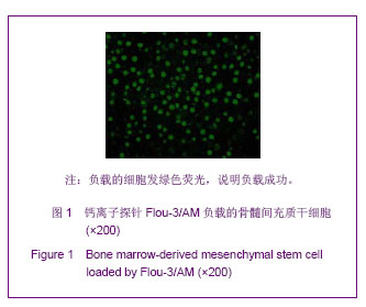
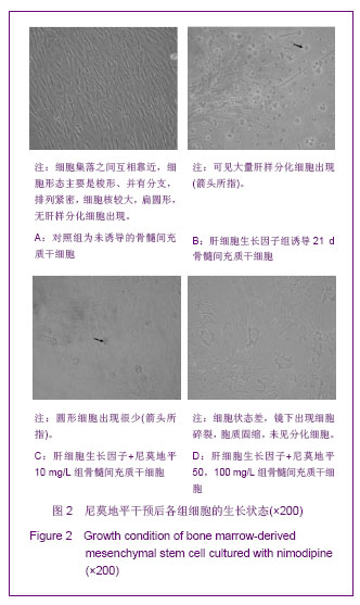
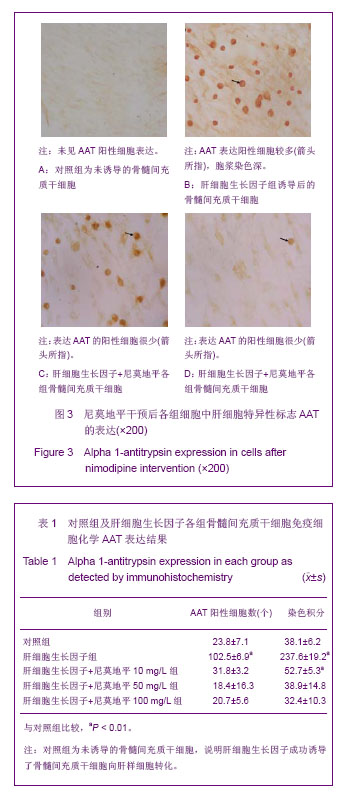
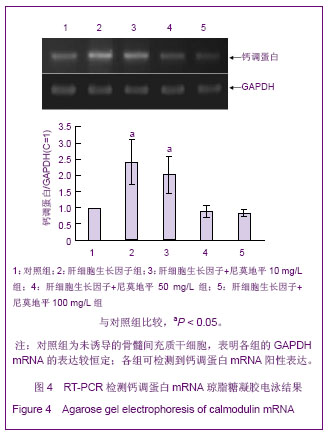
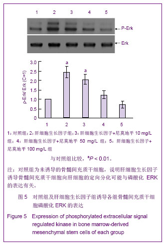
背景:骨髓间充质干细胞增殖和分化中的具体机制仍不清楚,Ca 2+信号、骨髓间充质干细胞增殖与分化信号如何协调和交叉成复杂信号网络等问题仍有待研究阐明。
目的:探讨细胞内Ca 2+在骨髓间充质干细胞向肝细胞定向诱导分化过程中的作用。
方法:用全骨髓贴壁法从大鼠骨髓中分离骨髓间充质干细胞后进行纯化和扩增,并加入肝细胞生长因子诱导骨髓间充质干细胞向肝细胞分化,应用流式细胞技术分别检测肝细胞生长因子诱导分化骨髓间充质干细胞和对照骨髓间充质干细胞内游离[Ca 2+ ]i。将不同浓度的尼莫地平加入肝细胞生长因子诱导的骨髓间充质干细胞(肝细胞生长因子组)培养液中进行干预后分为3组:肝细胞生长因子+尼莫地平10 mg/L组、肝细胞生长因子+尼莫地平50 mg/L组、肝细胞生长因子+尼莫地平100 mg/L组,在倒置相差显微镜下观察细胞生长情况并用免疫细胞化学法检测AAT的表达;并用RT-PCR检测对照组和尼莫地平干预组钙调蛋白mRNA的表达,免疫印迹法检测上述各组磷酸化ERK的表达。
结果与结论: ①加入肝细胞生长因子诱导分化的骨髓间充质干细胞内[Ca 2+]i显著高于对照组(P < 0.05)。②加入较大剂量的尼莫地平干预后,未见分化细胞且骨髓间充质干细胞的生长状态差;肝细胞生长因子+尼莫地平各组表达AAT的阳性细胞很少。③与对照组比较,肝细胞生长因子组和肝细胞生长因子+尼莫地平10 mg/L组钙调蛋白mRNA表达显著增加了(P < 0.05),肝细胞生长因子+尼莫地平50 mg/L组、肝细胞生长因子+尼莫地平100 mg/L组与对照组比较组间差异无显著性意义(P > 0.05)。说明Ca 2+不仅参与细胞因子诱导骨髓间充质干细胞向肝细胞的定向分化,而且也参与维持骨髓间充质干细胞的存活和增殖。

关键词: 干细胞, 骨髓干细胞, 骨髓间充质干细胞, Ca 2+ , 肝细胞生长因子, 细胞因子, 细胞分化, ERK, 干细胞图片文章

Abstract:

BACKGROUND: The mechanism of differentiation and proliferation of bone marrow-derived mesenchymal stem cells remains unclear. In addition, issues such as how signal pathways such as Ca 2+and bone marrow-derived mesenchymal stem cell proliferation and differentiation signals form complex signal network remain poorly understood.
OBJECTIVE: To investigate the effect of Ca 2+ in the induced differentiation of bone marrow-derived mesenchymal stem cells into hepatocytes.
METHODS: Bone marrow-derived mesenchymal stem cells were isolated from rat bone marrow using whone bone marrow adherence method, purified, amplified, and induced with hepatocyte growth factor. [Ca 2+ ]i in the  directional differentiated bone marrow-derived mesenchymal stem cells and control bone marrow-derived mesenchymal stem cells were detected with flow cytometry. Bone marrow-derived mesenchymal stem cells induced with hepatocyte growth factor were mixed with nimodipine of different concentration, and cells were divided into three groups: hepatocyte growth factor+ nimodipine 10 mg/L, 50 or 100 mg/L groups. Cell growth was observed with inverted phase contrast microscope and alpha 1-antitrypsin expression of the cells was confirmed by immunocytochemistry. The calcineurin M and the activation of extracellular signal regulated kinase pathway was detected by reverse transcription-PCR and western blotting, respectively.
RESULTS AND CONCLUSION: [Ca 2+ ]i in the directional differentiated bone marrow-derived mesenchymal stem cells was higher than in the control group (P < 0.05). After addition of a larger dose of nimodipine, no differentiation of cells was obeserved and growth of bone marrow-derived mesenchymal stem cells was getting worse. There were few alpha 1-antitrypsin positive cells in the nimodipine groups. Calcineurin M expression was significantly increased in directional differentiated bone marrow-derived mesenchymal stem cells and small dose of nimodipine than the controls (P < 0.05). However, no significant difference was found among middle, high dose nimodipine and control groups (P > 0.05). These findings indicate that Ca 2+ could participate in the differentiation of bone marrow-derived mesenchymal stem cells into hepatocytes incuded with cytokines, and also maintain the survival and proliferation of bone marrow-derived mesenchymal stem cells.

Key words: stem cells, mesenchymal stem cells, hepatocyte growth factor, cell differentiation

中图分类号: