Design
A randomized controlled animal experiment.
Time and setting
The experiment was conducted in the Second Hospital of Shandong University between January 2011 and April 2012.
Materials
Thirty-three SPF healthy Sprague-Dawley rats weighing 200-250 g were purchased from the Animal Experimental Center of Shandong University. Animal license was SCXK(Lu)20030004. During the experiment, the free drink, feeding feed and mixed feed were provided by Shandong University Animal Experiment Center, the room temperature was 18- 25 ℃, relative humidity was 62%-72%. The study protocol was reviewed and approved by the Institutional Animal Care and Use Committee, the Second Hospital of Shandong University, and the experiments were conducted according to the Guidelines of the American Physiological Society.
Chemicals and reagents are as follows:
.jpg)
Methods
Establishment and evaluation of pulmonary arterial hypertension model
Monocrotaline was prepared as described and was dissolved in 1 mol/L HCl, neutralized to pH 7.4 with
0.5 mol/L NaOH, and diluted with saline just before injection[2]. One week after monocrotaline injection[5], the rats were anesthetized and inserted with a 3F-Miller microtip catheter via the right jugular vein into the right ventricle to obtain baseline measurements of hemodynamics, such as right ventricular systolic pressure, mean right ventricular pressure, and mean pulmonary arterial pressure. The rats were euthanized after hemodynamic measurements, the lung and heart were quickly harvested and fixed in situ via the trachea cannula with buffered 4% formaldehyde, and then were embedded in paraf?n. The sections were cut into 4-
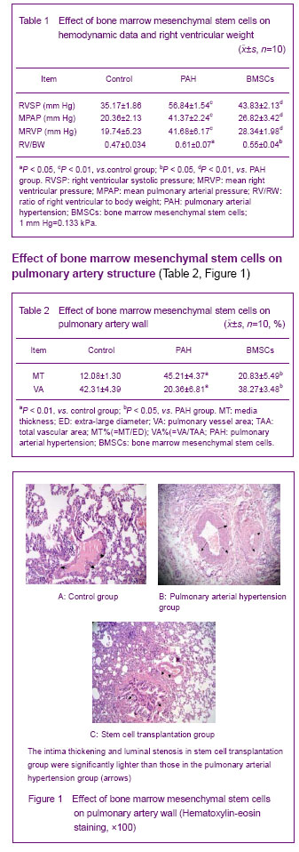
5 μm slices and were stained with streptavidin peroxidase and hematoxylin-eosin. According to the results of hemodynamic parameters, right ventricular hypertrophy and pulmonary arterial pathological changes were used to evaluate if the model of pulmonary arterial hypertension was successful establishment.
Isolation, culture and labeling of bone marrow mesenchymal stem cells
Bone marrow cells were isolated by flushing the cavity of femurs and tibias and transferred to a tissue culture dish with 90 mm in diameter. The bone marrow mesenchymal stem cells and other cells were separated with the ficoll (1.077) density gradient centrifugation method as previously report[6].
Flow cytometry immunophenotyping was performed by using methods reported previously: 5×10⁵ cells were suspended with trypsin and washed two times in PBS. After centrifugation, the cells were incubated with primary against human CD44, CD29, CD34, and CD90 antibodies for 30 minutes at 4 ℃.
Before implantation, the cells were labeled with the cross-linkable membrane dye
Cells were labeld according to the protocol of the supplier as previously described[7]. Briefly, (1-5)×105 cells were incubated for 5 minutes at 37 ℃, and then for an additional 15 minutes at 4 ℃.
After labeling, 1×10⁵ cells were being washed with PBS and resuspended in 100 μL saline and then kept on ice before transplantation, the labeling-efficiency reached more than 80%.
Groups of experimental animals
The male Sprague Dawley rats were randomly divided into three groups (n=10) as follows. Control group: animals received a sublingual vein injection of 0.9% saline instead of bone marrow mesenchymal stem cells. Pulmonary arterial hypertension group: animals received a subcutaneous injection of 50 mg/kg monocrotaline. Bone marrow mesenchymal stem cells group: animals received a sublingual vein injection of (1-5)×105 labeled bone marrow mesenchymal stem cells.
Evaluation of hemodynamic parameter and right ventricular impairment induced by monocrotaline
Two weeks after stem cell transplantation, the rats were anesthetized and inserted with a 3F-Miller microtip catheter via the right jugular vein into the right ventricle to obtain base line measurements of hemodynamics such as right ventricular systolic pressure, mean right ventricular pressure, and mean pulmonary arterial pressure[8]. The ratio of right ventricular to body weight was determined to measure the right ventricular hypertrophy[9].
Analysis of pulmonary vascular structural change
The morphometric analysis of pulmonary arteries was performed as described previously[10]. The structural changes in pulmonary vascular wall were observed by microscope, and the media thickness, extra-large diameter, pulmonary vessel area, and total vascular area were measured to calculate the ratio of media thickness/ extra-large diameter (%) and pulmonary vessel area/total vascular area (%). The ratio of right ventricular to body weight was determined to measure the right ventricular hypertrophy.
Analysis of cell differentiation by immunohistochemistry
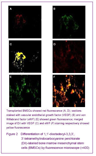
Two weeks after cell transplantation, the rats were anesthetized and the lung was inflated with optimal cutting temperature compound, and then quickly frozen in liquid nitrogen and stored at -80 ℃. Sections were cut into 4 μm slices and fixed in acetone for 10 minutes at -20 ℃. Survival of mesenchymal stem cells was demonstrated by observing the presence of
1, 1’-dioctadecyl-3, 3, 3’, 3’- tetramethylindocarbocyanine perchlorate(DiI)-labeled cells. Immunofluorescence was then carried out with goat anti-mouse monoclonal surfactant associated protein C (1:100) IgG antibody, rabbit anti-human von Willebrand factor (1:100) antibody and vascular endothelial growth factor (1:100) anti-body. Fluorescein isothiocyanate-conjugated goat anti-mouse IgG antibody was used as a secondary antibody. Two weeks after bone marrow mesenchymal stem cell implantation, the labeled red fluorescence-positive cells were observed by fluorescence microscope. The engraftment and integration of transplanted undifferentiated cells were identified through observing the distribution of red fluorescent-positive cells. The sections stained with von Willebrand factor, then the vascular endothelial growth factor and surfactant associated protein C presented as green fluorescence, and the merged images staining were yellow color.
Main outcome measures
The analysis of animal determination of hemodynamic data; right ventricular weigh and pulmonary vascular structural induced by monocrotaline; characterization of cultured bone marrow mesenchymal stem cells; the identification of the transplanted bone marrow mesenchymal stem cells; effect of bone marrow mesenchymal stem cells on the hemodynamic data and right ventricular impairment; the effect of bone marrow mesenchymal stem cells on pulmonary artery wall.
Statistical analysis
Statistical analysis was performed with one-way analysis of variance followed by Bonferroni test or t-test when appropriate by using SPSS 13.0 statistical software. Differences were considered significant at P < 0.05, and presented as mean±SD unless otherwise stated.