中国组织工程研究 ›› 2013, Vol. 17 ›› Issue (36): 6388-6395.doi: 10.3969/j.issn.2095-4344.2013.36.002
• 骨髓干细胞 bone marrow stem cells • 上一篇 下一篇
骨髓间充质干细胞的分化与静水压力刺激强度和持续时间
何 川1,2,梁 静2,邓廉夫2,冯建民1
- 1上海交通大学医学院附属瑞金医院骨科,上海市 200025;2上海市伤骨科研究所,上海市 200025
-
收稿日期:2013-02-19修回日期:2013-03-20出版日期:2013-09-03发布日期:2013-09-03 -
通讯作者:邓廉夫,博士,研究员,教授。上海市伤骨科研究所,上海市 200025 lfdeng@msn.com -
作者简介:何川☆,男,1971年生,四川省泸州市人,汉族,2005年上海第二医科大学毕业,博士,副主任医师,主要从事关节外科、人工关节的研究。 drhechuan@sina.com -
基金资助:国家自然科学基金项目(81071473)*;上海交通大学医学院自然科学研究基金项目(2008XJ038)*
Correlation between magnitude and duration of hydrostatic pressure and the differentiation of human bone marrow mesenchymal stem cells
He Chuan1, 2, Liang Jing2, Deng Lian-fu2, Feng Jian-min1
- 1 Department of Orthopedics, Ruijin Hospital, Shanghai Jiao Tong University School of Medicine, Shanghai 200025, China; 2 Shanghai Institute of Traumatology and Orthopaedics, Shanghai 200025, China
-
Received:2013-02-19Revised:2013-03-20Online:2013-09-03Published:2013-09-03 -
Contact:Deng Lian-fu, M.D., Researcher, Professor, Shanghai Institute of Traumatology and Orthopaedics, Shanghai 200025, China lfdeng@msn.com -
About author:He Chuan☆, M.D., Associate chief physician, Department of Orthopedics, Ruijin Hospital, Shanghai Jiao Tong University School of Medicine, Shanghai 200025, China; Shanghai Institute of Traumatology and Orthopaedics, Shanghai 200025, China drhechuan@sina.com -
Supported by:National Natural Science Foundation of China, No. 81071473*; Natural Science Foundation of Shanghai Jiao Tong University School of Medicine, No. 2008XJ038*
摘要:
背景:力学信号与骨骼系统的生长发育、修复重建和疾病发生发展有密切联系,其对骨髓间充质干细胞的影响和作用机制值得关注。 目的:探讨静水压力刺激对骨髓间充质干细胞分化的影响及相关机制。 方法:①短期实验:将人骨髓间充质干细胞分别接种于正常DMEM培养基、成骨诱导培养基或含细胞外信号调节激酶1/2抑制剂U0126的DMEM培养基中,利用自制压力加载系统对其施加0,40,80 kPa的静水压强1,4 h。②长期实验:将人骨髓间充质干细胞分别接种于正常DMEM培养基或成骨诱导培养基,施加40 kPa的静水压强4 h/d,持续14 d,以不施加静水压的细胞作对照。 结果与结论:实时定量反转录PCR结果显示,经成骨诱导或40 kPa静水压刺激 4 h后,骨髓间充质干细胞内核心结合因子α1、骨钙素mRNA表达增加,过氧化物酶体增殖物活化受体γ2和脂肪酶mRNA表达降低,80 kPa静水压刺激没有出现这一规律;40 kPa静水压的成骨诱导作用可被U0126部分拮抗。组织化学染色显示40 kPa静水压刺激7 d,骨髓间充质干细胞碱性磷酸酶表达和活性均增加;持续刺激14 d,过氧化物酶体增殖物活化受体γ2和脂肪酶mRNA表达增加。说明一定强度和作用时间的静水压刺激可调节骨髓间充质干细胞分化,其作用部分依赖于细胞外信号调节激酶1/2信号途径。
中图分类号:
引用本文
何 川,梁 静,邓廉夫,冯建民. 骨髓间充质干细胞的分化与静水压力刺激强度和持续时间[J]. 中国组织工程研究, 2013, 17(36): 6388-6395.
He Chuan, Liang Jing, Deng Lian-fu, Feng Jian-min. Correlation between magnitude and duration of hydrostatic pressure and the differentiation of human bone marrow mesenchymal stem cells[J]. Chinese Journal of Tissue Engineering Research, 2013, 17(36): 6388-6395.
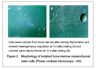
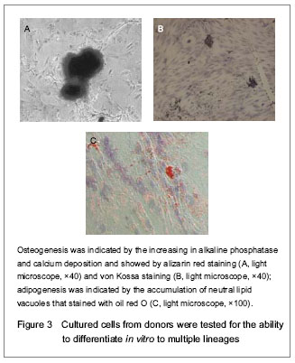
Multipotential of isolated human bone marrow mesenchymal stem cellsThe differentiation potential of the human bone marrow mesenchymal stem cells from three adult donors was observed by culturing the cells under conditions that favorable for adipogenic or osteogenic differentiation. After 1-3 weeks in lineage-specific culture conditions, the expanded cells were highly differentiated (the phenotype of lineage-specific cell types was easily distinguished) without evidence of the other lineages (Figure 3).
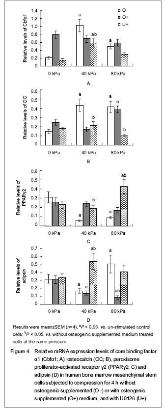
Effects of short-time hydrostatic pressure on the differentiation of human bone marrow mesenchymal stem cells Human bone marrow mesenchymal stem cells cultured in Dulbecco’s modified Eagle’s medium and stimulated with 40 kPa pressure for 4 hours showed a significant increasing in the expression of the osteogenic marker gene of core binding factor α1 and osteocalcin, and a significant decreasing in the expressions of the adipogenic marker genes of peroxisome proliferator- activated receptor γ2 and adipsin, when compared with controls (P < 0.05). Human bone marrow mesenchymal stem cells subjected to 80 kPa pressure for 4 hours showed a decreasing in core binding factor α1 expression (P < 0.05). Except for a decreasing in adipsin expression, there were no significant differences between cells cultured in osteogeneic supplemented medium in the presence of hydrostatic pressure and control cells. When extracellular signal-regulated kinase 1/2 activity was blocked by U0126, a significant decreasing in core binding factor α1 expression was observed in cells pressurized with 40 kPa for 4 hours, although core binding factor α1 expression was still higher than that in the non-pressurized control cells. Furthermore, pressurized cells with extracellular signal-regulated kinase 1/2 inhibition showed a decreasing in osteocalcin expression, reaching levels of expression showed no statistically significant difference from that of nonpressurized control cells. In contrast, pressurized cells with extracellular signal-regulated kinase 1/2 inhibition showed a significant increasing of peroxisome proliferator-activated receptor γ2 expression and adipsin expression (Figure 4).
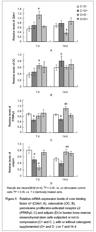
Effects of long-term hydrostatic pressure on the differentiation of human bone marrow mesenchymal stem cells (Figure 5) Hydrostatic pressure of 40 kPa for 4 hours per day could promote the expression of the osteogenic marker genes core binding factor α1 and osteocalcin after 7 days. Interestingly, the same hydrostatic pressure conditions continued for up to 14 days had a very different effect on cells, it could increase the expressions of adipogenic marker genes of peroxisome proliferator-activated receptor γ2 and adipsin. There was no synergistic effect under hydrostatic pressure and osteogenic induction.
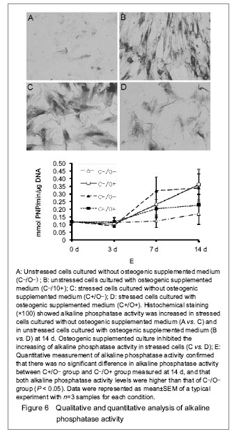
Qualitative and quantitative analysis of alkaline phosphatase after long-term hydrostatic pressure stressHydrostatic pressure or osteogenic supplemented medium alone could increase the activity and expression of alkaline phosphatase in human bone marrow mesenchymal stem cells, but this effect was not observed when two stimulus combined (Figure 6).
| [1] Costa P, Almeida FV, Connelly JT. Biophysical signals controlling cell fate decisions: how do stem cells really feel? Int J Biochem Cell Biol. 2012;44(12):2233-2237.
[2] Huang C, Ogawa R. Effect of hydrostatic pressure on bone regeneration using human mesenchymal stem cells. Tissue Eng Part A. 2012;18(19-20):2106-2113.
[3] Delaine-Smith RM, MacNeil S, Reilly GC. Matrix production and collagen structure are enhanced in two types of osteogenic progenitor cells by a simple fluid shear stress stimulus. Eur Cell Mater. 2012;24:162-174.
[4] Mathieu PS, Loboa EG. Cytoskeletal and focal adhesion influences on mesenchymal stem cell shape, mechanical properties, and differentiation down osteogenic, adipogenic, and chondrogenic pathways. Tissue Eng Part B Rev. 2012; 18(6):436-444.
[5] Milan JL, Lavenus S, Pilet P, et al. Computational model combined with in vitro experiments to analyse mechanotransduction during mesenchymal stem cell adhesion. Eur Cell Mater. 2013;25:97-113.
[6] Jaiswal RK, Jaiswal N, Bruder SP, et al. Adult human mesenchymal stem cell differentiation to the osteogenic or adipogenic lineage is regulated by mitogen-activated protein kinase. J Biol Chem. 2000;275(13):9645-9652 .
[7] Simmons CA, Matlis S, Thornton AJ, et al. Cyclic strain enhances matrix mineralization by adult human mesenchymal stem cells via the extracellular signal-regulated kinase (ERK1/2) signaling pathway. J Biomech. 2003;36(8):1087-1096.
[8] Thompson WR, Rubin CT, Rubin J. Mechanical regulation of signaling pathways in bone. Gene. 2012; 503(2):179-193.
[9] Wenisch S, Trinkaus K, Hild A, et al. Human reaming debris: a source of multipotent stem cells. Bone. 2005;36(1):74-83.
[10] Lee HS, Huang GT, Chiang H, et al. Multipotential mesenchymal stem cells from femoral bone marrow near the site of osteonecrosis. Stem Cells. 2003;21(2):190-199.
[11] He C, Liang J, Deng LF, et al. Effect of hydrostatic compressive loading on the cytoskeletal structure of human bone marrow mesenchymal stem cells. Zhongguo Zuzhi Gongcheng Yanjiu yu Linchuang Kangfu. 2011;15(45): 8366-8370.
[12] Robertson N, Leek R. Isolation of RNA from tumor samples: single-step guanidinium acid-phenol method. Methods Mol Med. 2006;120:55-9.
[13] Liu J, Zhao Z, Li J, et al. Hydrostatic pressures promote initial osteodifferentiation with ERK1/2 not p38 MAPK signaling involved. J Cell Biochem. 2009;107(2):224-232.
[14] Glossop JR, Cartmell SH. Effect of fluid flow-induced shear stress on human mesenchymal stem cells: differential gene expression of IL1B and MAP3K8 in MAPK signaling. Gene Expr Patterns. 2009;9(5):381-388.
[15] Verma D, Ye N, Meng F, et al. Interplay between cytoskeletal stresses and cell adaptation under chronic flow. PLoS One. 2012;7(9):e44167.
[16] Steward AJ, Wagner DR, Kelly DJ. The pericellular environment regulates cytoskeletal development and the differentiation of mesenchymal stem cells and determines their response to hydrostatic pressure. Eur Cell Mater. 2013;25:167-178.
[17] Ziros PG, Gil AP, Georgakopoulos T, et al. The bone-specific transcriptional regulator Cbfa1 is a target of mechanical signals in osteoblastic cells. J Biol Chem. 2002;277(26):23934-23941.
[18] Liu J, Zou L, Wang J, et al. Hydrostatic pressure promotes Wnt10b and Wnt4 expression dependent and independent on ERK signaling in early-osteoinduced MSCs. Biochem Biophys Res Commun. 2009;379(2):505-509. |
| [1] | 姜 涛, 马 磊, 李志强, 寿 玺, 段明军, 吴 硕, 马 创, 魏 琴. 血小板衍生生长因子BB诱导骨髓间充质干细胞向血管内皮细胞分化[J]. 中国组织工程研究, 2021, 25(25): 3937-3942. |
| [2] | 陈 扬, 黄邓高, 高元慧, 王顺兰, 曹 卉, 郑琳麟, 何浩伟, 罗思琴, 肖敬川, 张应爱, 张淑芳. 低强度脉冲场超声促进人脂肪间充质干细胞的增殖和黏附[J]. 中国组织工程研究, 2021, 25(25): 3949-3955. |
| [3] | 张立书, 刘安琪, 何小宁, 金 岩, 李 蓓, 金 钫. Alpl基因影响骨髓间充质干细胞治疗溃疡性结肠炎[J]. 中国组织工程研究, 2021, 25(25): 3970-3975. |
| [4] | 阮光萍, 姚 翔, 刘高米洋, 蔡学敏, 李自安, 庞荣清, 王金祥, 潘兴华. 脐带间充质干细胞移植治疗树鼩创伤性全身炎症反应综合征[J]. 中国组织工程研究, 2021, 25(25): 3994-4000. |
| [5] | 莫剑玲, 何少茹, 冯博文, 简敏桥, 张晓晖, 刘财盛, 梁一晶, 刘玉梅, 陈 亮, 周海榆, 刘艳辉. 构建预血管化细胞膜片及血管形成相关因子的表达[J]. 中国组织工程研究, 2021, 25(22): 3479-3486. |
| [6] | 陈 磊, 郑 蕊, 杰永生, 綦 惠, 孙 磊, 舒 雄. 脂肪血管基质成分复合骨软骨一体化支架的体外评价[J]. 中国组织工程研究, 2021, 25(22): 3487-3492. |
| [7] | 魏 琴, 张 雪, 马 磊, 李志强, 寿 玺, 段明军, 吴 硕, 贾麒钰, 马 创. 血小板衍生生长因子BB诱导大鼠骨髓间充质干细胞向成骨细胞分化[J]. 中国组织工程研究, 2021, 25(19): 2953-2957. |
| [8] | 陈 晓, 郭 智, 陈丽娜, 刘玄勇, 张弋慧智, 李旭绵, 王月乔, 韦丽娅, 谢 晶, 蔺 莉. 自体外周血造血干细胞动员采集的影响因素[J]. 中国组织工程研究, 2021, 25(19): 2958-2962. |
| [9] | 郭志斌, 吴春芳, 刘子洪, 张钰英, 迟博婧, 王 宝, 马 超, 张国彬, 田发明. 辛伐他汀可刺激骨髓间充质干细胞的成骨分化[J]. 中国组织工程研究, 2021, 25(19): 2963-2968. |
| [10] | 李聪聪, 姚 楠, 黄丹娥, 宋 敏, 彭 莎, 李安安, 卢 超, 刘文刚. 人髌下脂肪垫干细胞的鉴定及成软骨分化[J]. 中国组织工程研究, 2021, 25(19): 2976-2981. |
| [11] | 高元慧, 向 杨, 曹 卉, 王顺兰, 郑琳麟, 何浩伟, 张应爱, 张淑芳, 黄邓高. 两种月龄近交系五指山小型猪脂肪间充质干细胞生物学特性的比较[J]. 中国组织工程研究, 2021, 25(19): 2988-2993. |
| [12] | 曹 阳, 张军平, 彭 立, 丁 义, 李光辉. 兔主动脉内皮细胞的分离培养及生物学特性[J]. 中国组织工程研究, 2021, 25(19): 3000-3003. |
| [13] | 代 敏, 王 帅, 张霓霓, 黄桂林, 余丽梅, 胡小华, 易 杰, 姚 礼, 张立刚. 低氧预处理人羊膜间充质干细胞的生物学特征[J]. 中国组织工程研究, 2021, 25(19): 3004-3008. |
| [14] | 覃艳春, 荣 震, 蒋锐沅, 付 彬, 洪晓华, 莫春梅. 中药复方制剂抑制CD133+肝癌干细胞增殖及干性转录因子的表达[J]. 中国组织工程研究, 2021, 25(19): 3016-3023. |
| [15] | 戴雅玲, 陈乐文, 何肖君, 林华伟, 贾微微, 陈立典, 陶 静, 柳维林. miR-146b过表达慢病毒载体构建及对海马神经干细胞增殖的影响[J]. 中国组织工程研究, 2021, 25(19): 3024-3030. |
Design: Cytology contrast study.
.jpg)
10 mmol/L U0126, which blocks the extracellular signal-regulated kinase 1/2 pathway. Cells were then immediately subjected for mechanical stimulation. Using a customized air pressure vessel (Figure 1), a sustained hydrostatic pressure of 40 or 80 kPa was applied to the cells for 1 or 4 hours. In the long-term study, a sustained hydrostatic pressure of 40 kPa was applied to human bone marrow mesenchymal stem cells (cultured in 10% Dulbecco’s modified Eagle’s medium or osteogenic supplemented medium) for 4 hours every day and lasted for 14 days. Control culture plates were setup in identical conditions, except that no pressure was applied.
.jpg)
1 mmol/L MgCl2, pH 10.5) at 37 ℃ for 10-20 minutes. The absorbance was measured with an ultraviolet spectrophotometer DU640 at 405 nm and converted to p-nitrophenol concentration based on standard solutions prepared in parallel. To determine the amount of DNA in each well, the cell nuclei were disrupted by addition of lysis buffer (0.025 mol/L tris-HCl, 0.4 mol/L NaCl, 0.5% SDS, pH 7.4), followed by sonication and centrifugation. The DNA content was determined from the supernatant using the Hoescht 33258 assay and standard solutions of calf thymus DNA prepared in parallel.
Independent experiments were performed at least four times in duplicate cultures (unless otherwise noted). Data were expressed as mean±SEM and analyzed for levels of significance with SPSS 11.5 using the Student’s t-test (one sample t-test and paired t-test, where appropriate). P value < 0.05 was considered as statistical significant.
1力学信号是肌肉骨骼组织生长发育的重要调控因素,在细胞水平的修复过程中力学信号也起重要的调控作用。 2骨髓间充质干细胞成骨/成脂分化调控涉及许多骨科疾患的病理演变过程,实验重点研究压应力信号对其影响。 3 实验结果发现,40 kPa静水压可以调节骨髓间充质干细胞分化,其作用部分依赖于细胞外信号调节激酶1/2信号途径。
Mechanical load, such as gravity and body posture, produces biomechanical stimulations in the bone tissue, and plays an important role in the process of bone development, growth, modeling and remodeling. The differentiation of mesencnymal stem cells and the progenitors of multiple cellular lineages in bone, may decide the quality, quantity, and structure of the new bone formation. Mesenchymal stem cells have been proven to be responsive to mechanical signals such as hydrostatic pressure and to shear stress induced by fluid flow in their environment. High intraosseous pressure, suggesting high hydrostatic pressure in bone, is usually occurs in the pathology of some bone diseases, such as osteonecrosis, osteoarthritis, and osteomyelitis. We demonstrated herein that hydrostatic pressure can influence the osteogenic and adipogenic differentiation of human bone marrow mesenchymal stem cells in vitro.
| 阅读次数 | ||||||
|
全文 |
|
|||||
|
摘要 |
|
|||||