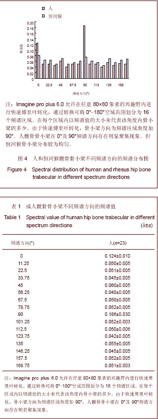
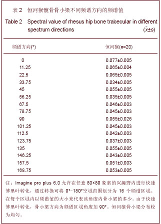
| [1] Wolff JL. The Law of Bone Remodeling. Berlin: Springer. 1986.[2] Bergmann G, Graichen F, Rohlmann A. Hip joint loading during walking and running, measured in two patients. J Biomech. 1993;26(8):969-990. [3] Weigel JP, Wasserman JF. Biomechanics of the normal and abnormal hip joint. Vet Clin North Am Small Anim Pract. 1992; 22(3):513-528. [4] Chan CW, Rudins A. Foot biomechanics during walking and running. Mayo Clin Proc. 1994;69(5):448-461.[5] Dalstra M, Huiskes R. Load transfer across the pelvic bone. J Biomech. 1995;28(6):715-724. [6] Heller MO, Schröder JH, Matziolis G, et al. Musculoskeletal load analysis. A biomechanical explanation for clinical results--and more? Orthopade. 2007;36(3):188, 190-194. [7] Sverdlova NS, Witzel U. Principles of determination and verification of muscle forces in the human musculoskeletal system: Muscle forces to minimise bending stress. J Biomech. 2010;43(3):387-396. [8] Dietrichs E. Anatomy of the pelvic joints--a review. Scand J Rheumatol Suppl. 1991;88:4-6. [9] Savvidis E. Stress on the coxal end of the femur during loading in the hip joint flexion posture. Aktuelle Probl Chir Orthop. 1990;39:1-56. [10] Leopold D, Novotny V. Sex determination from the skull and parts of the hip bone. Gegenbaurs Morphol Jahrb. 1985; 131(3): 277-285. [11] Leutenegger W. The pelvis of the recent primates. Gegenbaurs Morphol Jahrb. 1970;115(1):1-101. [12] Dietrichs E, Hansen JH, Bakland O. Anatomy of the pelvic joints. Tidsskr Nor Laegeforen. 1990;110(17):2213-2215.[13] Rook L, Bondioli L, Köhler M, et al. Oreopithecus was a bipedal ape after all: evidence from the iliac cancellous architecture. Proc Natl Acad Sci U S A. 1999;96(15): 8795-8799.[14] Oxnard CE. Bone and bones, architecture and stress, fossils and osteoporosis. J Biomech. 1993;26 Suppl 1:63-79.[15] Pete E. Fourier Descriptors and Their Applications in Biology. Cambridge: Cambridge University Press 1997.[16] Watkins C, Sadun A. Marenka S. Modern Image Processing. Boston: Academic Press. 1993.[17] Anderson AE, Peters CL, Tuttle BD, et al. Subject-specific finite element model of the pelvis: development, validation and sensitivity studies. J Biomech Eng. 2005;127(3):364-373. [18] van Rietbergen B. Micro-FE analyses of bone: state of the art. Adv Exp Med Biol. 2001;496:21-30. [19] Knothe Tate ML, Tami AE, Netrebko P, et al. Multiscale computational and experimental approaches to elucidate bone and ligament mechanobiology using the ulna-radius-interosseous membrane construct as a model system. Technol Health Care. 2012;20(5):363-378. [20] Zhang M, Mak AF, Roberts VC. Finite element modelling of a residual lower-limb in a prosthetic socket: a survey of the development in the first decade. Med Eng Phys. 1998;20(5): 360-373. [21] Daegling DJ, Hylander WL. Experimental observation, theoretical models, and biomechanical inference in the study of mandibular form. Am J Phys Anthropol. 2000;112(4): 541-551.[22] Kristen KH, Berger K, Berger C, et al. The first metatarsal bone under loading conditions: a finite element analysis. Foot Ankle Clin. 2005;10(1):1-14. [23] Ross CF. Finite element analysis in vertebrate biomechanics. Anat Rec A Discov Mol Cell Evol Biol. 2005;283(2):253-258. [24] Richmond BG, Wright BW, Grosse I, et al. Finite element analysis in functional morphology. Anat Rec A Discov Mol Cell Evol Biol. 2005;283(2):259-274.[25] van der Meulen MC. Diaphyseal bone growth and adaptation: models and data. Stud Health Technol Inform. 1997;40:17-23. [26] Anderson AE, Ellis BJ, Weiss JA. Verification, validation and sensitivity studies in computational biomechanics. Comput Methods Biomech Biomed Engin. 2007;10(3):171-184. [27] Park HK, Dujovny M, Park T, et al. Application of finite element analysis in neurosurgery. Childs Nerv Syst. 2001; 17(1-2):87-96. [28] Henninger HB, Reese SP, Anderson AE, et al. Validation of computational models in biomechanics. Proc Inst Mech Eng H. 2010;224(7):801-812.[29] Dalstra M, Huiskes R, van Erning L. Development and validation of a three-dimensional finite element model of the pelvic bone. J Biomech Eng. 1995;117(3):272-278. [30] Mellal A, Wiskott HW, Botsis J, et al. Stimulating effect of implant loading on surrounding bone. Comparison of three numerical models and validation by in vivo data. Clin Oral Implants Res. 2004;15(2):239-248.[31] Turner CH, Cowin SC, Rho JY, et al. The fabric dependence of the orthotropic elastic constants of cancellous bone. J Biomech. 1990;23(6):549-561. [32] Odgaard A. Three-dimensional methods for quantification of cancellous bone architecture. Bone. 1997;20(4):315-328. [33] Katz JL, Meunier A. The elastic anisotropy of bone. J Biomech. 1987;20(11-12):1063-1070.[34] Nicholson PF. Ultrasound and the biomechanical competence of bone. IEEE Trans Ultrason Ferroelectr Freq Control. 2008; 55(7):1539-1545. [35] Wear KA. Ultrasonic scattering from cancellous bone: a review. IEEE Trans Ultrason Ferroelectr Freq Control. 2008; 55(7):1432-1441.[36] Gibson LJ. The mechanical behaviour of cancellous bone. J Biomech. 1985;18(5):317-328. Katz JL. [37] The structure and biomechanics of bone. Symp Soc Exp Biol. 1980;34:137-168. [38] Sharir A, Barak MM, Shahar R. Whole bone mechanics and mechanical testing. Vet J. 2008;177(1):8-17. [39] Jacobs CR. The mechanobiology of cancellous bone structural adaptation. J Rehabil Res Dev. 2000;37(2): 209-216. [40] Weiner S, Traub W, Wagner HD. Lamellar bone: structure-function relations. J Struct Biol. 1999;126(3): 241-255. [41] Paris O, Zizak I, Lichtenegger H, et al. Analysis of the hierarchical structure of biological tissues by scanning X-ray scattering using a micro-beam. Cell Mol Biol (Noisy-le-grand). 2000;46(5):993-1004. [42] Doblaré M, García JM. On the modelling bone tissue fracture and healing of the bone tissue. Acta Cient Venez. 2003;54(1): 58-75. [43] Burger EH, Klein-Nulen J. Responses of bone cells to biomechanical forces in vitro. Adv Dent Res. 1999;13:93-98.[44] Athanasiou KA, Zhu C, Lanctot DR, et al. Fundamentals of biomechanics in tissue engineering of bone. Tissue Eng. 2000;6(4):361-381. [45] Skerry TM, Suva LJ. Investigation of the regulation of bone mass by mechanical loading: from quantitative cytochemistry to gene array. Cell Biochem Funct. 2003;21(3):223-229.[46] Natali AN, Meroi EA. A review of the biomechanical properties of bone as a material. J Biomed Eng. 1989;11(4): 266-276. [47] Bruce DM. Mathematical modelling of the cellular mechanics of plants. Philos Trans R Soc Lond B Biol Sci. 2003;358 (1437): 1437-1444. [48] Klein-Nulend J, Bacabac RG, Mullender MG. Mechanobiology of bone tissue. Pathol Biol (Paris). 2005;53(10):576-580. [49] Barak MM, Sharir A, Shahar R. Optical metrology methods for mechanical testing of whole bones. Vet J. 2009;180(1):7-14. [50] Nyman JS, Reyes M, Wang X. Effect of ultrastructural changes on the toughness of bone. Micron. 2005;36(7-8): 566-582. [51] Frost HM. Vital biomechanics: proposed general concepts for skeletal adaptations to mechanical usage. Calcif Tissue Int. 1988;42(3):145-156. [52] Thwaites JJ, Mendelson NH. Mechanical behaviour of bacterial cell walls. Adv Microb Physiol. 1991;32:173-222. [53] Martinko V, Belay M, Jelínek L. Fundamentals of the biomechanics of hard tissues. Anat Anz. 1991;173(4): 225-232. [54] Iida M. Methods of determining the mechanical properties of bone. Nihon Seikeigeka Gakkai Zasshi. 1991;65(4):240-249. [55] Ito H. Mechanical stress for the bone and soft tissues. J Nippon Med Sch. 2002;69(2):146-148.[56] Hollinshead W, William H. Functional Anatomy of the Limbs and BAck. Philadelphia: Saunders. 1981. [57] Pedersen DR, Brand RA, Davy DT. Pelvic muscle and acetabular contact forces during gait. J Biomech. 1997;30(9): 959-965.[58] Thorpe SK, Crompton RH, Günther MM, et al. Dimensions and moment arms of the hind- and forelimb muscles of common chimpanzees (Pan troglodytes). Am J Phys Anthropol. 1999;110(2):179-199.[59] Dalstra M, Huiskes R, Odgaard A, et al. Mechanical and textural properties of pelvic trabecular bone. J Biomech. 1993;26(4-5):523-535. |
.jpg)
.jpg)

.jpg)
.jpg)