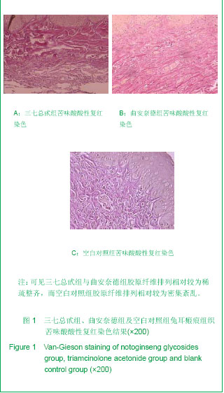
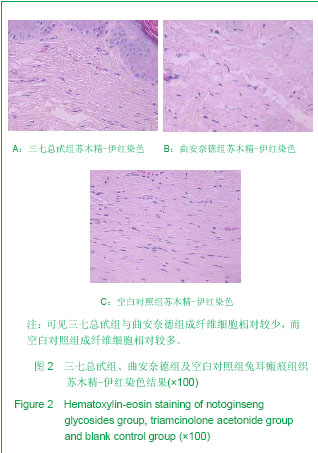
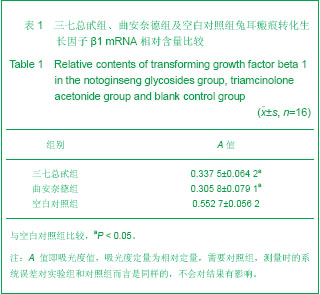
| [1] Bayat A, Bock O, Mrowietz U,et al. Genetic susceptibility to keloid diseaseand hypertrophic scaring:transforming growth factor β1 commonpolymorphisms and plasma levels. Plast Reconstr Surg. 2003;111:535-543.http://www.ncbi.nlm.nih.gov/pubmed/?term=Genetic+susceptibility+to+keloid+diseaseand+hypertrophic+scaring%3Atransforming+growth+factor+%CE%B2+1+commonpolymorphisms+and+plasma+levels. [2] Lekic P, McCulloch CA. Periodontal ligament cell populations : the centralrole of fibroblasts in creating a unique tissue. Anat Rec. 1996; 245(2):327-341.http://www.ncbi.nlm.nih.gov/pubmed/?term=Periodontal+ligament+cell+populations+%3A+the+centralrole+of+fibroblasts+in+creating+a+unique+tissue. [3] Amadeu TP, Coulomb B, Desmouliere A.Cutaneous wound healing:myofibroblasts differentiation and in vitro models. Lower Extrem Wounds. 2003; 2(2): 60-68.http://www.ncbi.nlm.nih.gov/pubmed/15866829 [4] Chen Y. Guangxi Yixue. 1998;20(6): 1109-1112.陈英. 三七总甙的的药理研究及临床应用进展[J]. 广西医学,1998, 20(6): 1109-1112.http://www.cqvip.com/Main/Detail.aspx?id=3305895 [5] Zhang RH. Disan Junyi Daxue Xuebao. 2000;22(4): 307-310.张荣华. 三七抗肝纤维化的实验研究[J]. 第三军医大学学报, 2000, 22(4): 307-310.http://doctor.51daifu.com/2007/0305/BB7565E7C948T119106.shtml [6] Dong XQ, Duan L, Pang LB, et al. Shandong Daxue Xuebao: Yixueban. 2012;50(1).85-88.董向前,段丽,平梁兵,等.人参皂苷 Rg1 和 Rb1 抗肝纤维化的体视学研究[J]. 山东大学学报: 医学版, 2012, 50(1).85-88.http://www.cnki.com.cn/Article/CJFDTotal-SDYB201201020.htm [7] Liu JY, Li SR, Ji SX, et al. Disan Junyi Daxue Xuebao. 2003;25(17);1562-1563.刘剑毅,李世荣,纪淑兴,等.三七总甙对人增生性瘢痕成纤维细胞增殖及胶原合成的作用[J].第三军医大学学报,2003,25(17);1562-1563.http://wuxizazhi.cnki.net/Search/DSDX200317026.html [8] Li HY. Xi′an: Fourth Military Medical University Press. 2005: 40-45.李荟元.创伤研究动物模型兔耳瘢痕模型的建立与应用[M]. 西安:第四军医大学出版社,2005: 40-45. http://www.bookschina.com/1082580.htm [9] Shu B, Hao LL, Wu ZY, et al. Disan Junyi Daxue Xuebao. 2001;23(3):343-345.舒彬,郝林林,吴宗耀, 等. 兔耳增生性瘢痕的形态学观察与羟脯氨酸含量变化[J].第三军医大学学报,2001, 23(3):343-345.http://d.wanfangdata.com.cn/Periodical_dsjydxxb200103029.aspx [10]Feng DC, Yang XM, Xue HB, et al. Zhongguo Meirong Yixue. 2009;18(2):191-194.冯登超,杨喜明,薛宏斌,等. 建立更加稳定和有效的兔耳瘢痕模[J]. 中国美容医学,2009,18(2):191-194.http://med.wanfangdata.com.cn/SearchCenter/searchInfomation.aspx?infoQuery=zgmryxzz200902021 [11]Yu R, Song Y, Chen JJ, et al. Huaxi Yixue. 2010;25(7):1209-1212.于蓉,宋烨,陈俊杰,等. 一种兔耳中期增生性瘢痕动物模型的建立[J].华西医学,2010,25(7):1209-1212.http://www.cqvip.com/Main/Detail.aspx?id=34957332 [12]Li H, Liu M, Chen Z, et al. Zhongguo Linchuang Yisheng Zazhi. 2007;35(6):45-46.李辉,刘民,陈织,等. 曲安奈德局部注射治疗瘢痕疙瘩60例疗效观察[J]. 中国临床医生杂志,2007,35(6):45-46.http://d.wanfangdata.com.cn/Periodical_zglcys200706022.aspx [13]Wang ZH, Wang DS, Gao GX. Zhongguo Yiyao Zhinan. 2010;8(12):88-89.王振华,王殿生,高国鑫.局部注射曲安奈德治疗耳垂瘢痕疙瘩[J]. 中国医药指南,2010,8(12):88-89.http://www.cqvip.com/QK/86373X/201012/33511680.html [14] Xie J, Wang Y, Zhang XL, et al. Linchuang Yixue yu Huli Yanjiu. 2006;5(6):16.谢俊,王宇,张晓龙, 等. 曲安奈德治疗增生性瘢痕的疗效观察[J]. 临床医学与护理研究, 2006,5(6):16.http://wuxizazhi.cnki.net/Search/AHWZ200606010.html [15] Yu R, Cen Y. Zhongguo Xiufu Chongjian Waike Zazhi. 2012;26(3):331-336.于蓉,岑瑛. TGF-β1/Smad3 信号转导通路与创伤后瘢痕形成[J]. 中国修复重建外科杂志,2012,26(3):331-336.http://www.doc88.com/p-691588606077.html [16]Abergei RP,Pizzurro D,Meeker CA.Biochemical composition of con-nective tissue in keloids and analysis of collagen metabolism in keloids fibroblast cultures.Invest Dermatol. 1985;84(5):384.http://www.ncbi.nlm.nih.gov/pubmed/?term=Biochemical+composition+of+con-nective+tissue+in+keloids+and+analysis+of+collagen+metabolism+in+keloids+fibroblast+cultures. [17]Wang X,Smith P,Pu LL,et al.Exogenous transforming growth factorβ2 modulates collagenⅠAnd col -lagenⅢsynthesis in proliferative scarxe-nografts in nude rats. J Surg Res. 1999;87(2):194-200.http://www.ncbi.nlm.nih.gov/pubmed/10600349 [18]Shah M,Foreman DM, Ferguson MW. Neutralisation of TGFβ1 and TGFβ2 or exogenous addition of TGFβ3 to cutaneous ratwoundsreduces scarring.J Cell Sci. 1995;108(3):985-986.http://www.ncbi.nlm.nih.gov/pubmed/7542672 [19] Shek FW, Benyon RC. How can transforming growth factorbe-ta be targeted usefully to combat liver fibrosis? Eur J Gastroenterol Hepatol. 2004;16(2): 127-133.http://www.ncbi.nlm.nih.gov/pubmed/?term=How+can+transforming+growth+factorbe-ta+be+targeted+usefully+to+combat+liver+fibrosis%3F [20]Cheng J, Grande JP. Transforming growth factorβsignaltransduction and progressive renal disease. Exp Biol Med(Maywood). 2002;227(6):943-956.http://www.ncbi.nlm.nih.gov/pubmed/12486204 [21]Grande JP,Warner GM,Walker HJ,et al.TGF-β1is an au-tocinesis mediator of renal tubule epithelial cell growth and col-lagen Ⅳ production. Exp Biol Med (Maywood).2002;227(3):171-181.http://www.ncbi.nlm.nih.gov/pubmed/11856815 [22]Ye X,Li H.A Study of Transforming growth factor β, vi-mentin and desmin gene expression in the renal cortex of dia-betic rats during the early diabetic period. J Zhejiang Univ:Med Sci. 2004;34(1):55-59.http://www.dissertationtopic.net/doc/1191941 [23]Goumenos DS,Tsamandas AC,Oldroy DS,et al.Transforming growth factor beta1 and myofibroblasts:a potential pathwaytowards renal scarring in human glomerular disease.Nephron. 2001;87(2):240-248.http://www.ncbi.nlm.nih.gov/pubmed/?term=Transforming+growth+factor+beta1+and+myofibroblasts%3Aa+potential+pathwaytowards+renal+scarring+in+human+glomerular+disease. [24]Yao H, Li SR, Liu JY. Zhongguo Shiyong Meirong Zhengxing Waike Zazhi. 2005;16(4):243-245.姚恒,李世荣,刘剑毅. 三七总甙对人增生性瘢痕成纤维细胞TGF-β1和细胞周期的作用[J].中国实用美容整形外科杂志, 2005,16(4):243-245.http://www.cnki.com.cn/Article/CJFDTotal-SMZW200504028.htm [25]Peng BG, Lv MD, Xiao DZ, et al. Zhongguo Yaolixue Tongbao. 1997; 13(1): 70-72.彭宝岗, 吕明德, 肖定璋, 等. 三七总皂甙对大鼠肝细胞钙内流的阻断作用[J]. 中国药理学通报, 1997, 13(1): 70-72.http://www.cqvip.com/Main/Detail.aspx?id=2566383 [26]Zhang R, Zhou ZC, Hong DL, et al. Disan Junyi Daxue Xuebao. 2002;22(4):307-310.张荣华,周子成,洪多伦,等.三七抗肝纤维化的实验研究[J].第三军医大学学报,2002,22(4):307-310.http://doctor.51daifu.com/2007/0305/BB7565E7C948T119106.shtml [27] Xiong L. Zhongcaoyao. 1998;29(8):567-568.熊磊.三七治疗肝病的实验研究进展[J].中草药,1998,29(8):567-568.http://2010.cqvip.com/qk/92286X/199808/3115591.html [28] Cheng H, Xiong L, Liu P, et al. Zhongguo Zhongxiyi Jiehe Piwei Zazhi. 1999;7(4):203-205.成海,熊磊,刘平,等. MTT法观察三七总甙对NIH/3T3成纤维细胞毒性作用与增殖的影响[J] 中国中西医结合脾胃杂志,1999, 7(4): 203-205.http://www.cnki.com.cn/Article/CJFDTotal-ZXPW199904005.htm [29]Shi XF, Xu M, Liu Q, et al. Zhongyao Yaoli yu Linchuang. 2010;17(2):7-8.石小枫,徐曼,刘杞, 等. 三七总皂甙对肝纤维化大鼠Ⅰ、Ⅲ胶原及TGF-β1的影响[J]中药药理与临床,2010,17(2):7-8.http://www.cnki.com.cn/Article/CJFDTotal-ZYYL200102004.htm [30]Wei Y, Fan JM, Pan LP, et al. Zhongguo Zhongxiyi Jiehe Zazhi. 2002;22(1): 47-49.韦颖,樊均明,潘丽萍,等.三七总皂甙对人肾成纤维细胞的影响[J].中国中西医结合杂志,2002,22(1): 47-49.http://www.cnki.com.cn/Article/CJFDTotal-ZZXJ200201022.htm [31]Zhang GQ, Ye RG, Kong QY, et al. Zhonghua Shenzangbing Zazhi. 1998;14(2):93-95.张国强,叶任高,孔庆瑜,等. 三七总甙诱导间质纤维化人肾成纤维细胞凋亡及其分子机理初探[J]. 中华肾脏病杂志, 1998, 14(2):93-95.http://www.chemdrug.com/databases/7_32_iivfdmehdxsdmmfk.html [32]Xu Q, Zhou W, Kong HY, et al. Zhongguo Gushang. 2010;23(4).278-281.徐泉,周卫,孔焕宇,等. 三七透明质酸钠凝胶对家兔椎板切除术后硬膜外瘢痕中胶原成分的影响[J].中国骨伤,2010,23(4).278-281.http://wuxizazhi.cnki.net/Search/ZGGU201004019.html [33] Yamasaki S,Nakashima T,Kawakami A,et al.Cytokines regulate fibroblast-like synovial cell differentiation to adipocyte-like cells.Rheumatology. 2004;43(4):448-452.http://www.ncbi.nlm.nih.gov/pubmed/?term=Cytokines+regulate+fibroblast-like+synovial+cell+differentiation+to+adipocyte-like+cells. [34]Tao JL, Guo M, Liu H, et al. Zhongguo Zhongxiyi Jiehe Shenbing Zazhi. 2008;9(9):799-801.陶静莉,郭敏,刘华,等.三七总苷对慢性肾衰竭大鼠模型肾纤维化的治疗作用及机制[J].中国中西医结合肾病杂志,2008,9(9):799-801.http://www.cqvip.com/qk/83620X/200809/28352394.html [35]Quan Y, Xia QM, Li FX, et al. Xinan Guofang Yiyao. 2009;19(1):23-25.全燕,夏前明,李福祥,等.三七总皂苷对大鼠肺纤维化及结缔组织生长因子表达的影响[J].西南国防医药,2009,19(1):23-25.http://www.cnki.com.cn/Article/CJFDTotal-XNGF200901015.htm [36]Zhang XW, Li FX, Li H. Zhongguo Xinyao Zazhi. 2007;16(4):260-262.张娴文,李凤贤,李辉. 三七皂苷抗肝纤维化作用的现状[J].中国新药杂志,2007,16(4):260-262.http://www.cnki.com.cn/Article/CJFDTotal-ZXYZ200704001.htm [37]Lv WH, Xu S, Zhang HQ. Zhongguo Yesheng Zhiwu Ziyuan. 2009;28(2):1-4.吕伟红,徐姗,张洪泉. 三七总皂苷抗肝纤维化作用机制的研究进展[J].中国野生植物资源,2009,28(2):1-4.http://wzhz.cqvip.com/hzpinglaoshi/qk/98203A/200801/26399819.html [38] Laxova A, Earley M, Farrell P, et al. Results of DNA-based cystic fibrosis newborn screening proficiency testing. J Cystic Fibrosis. 2008;6(2): S14.http://www.ncbi.nlm.nih.gov/pubmed/?term=Results+of+DNA-based+cystic+fibrosis+newborn+screening+proficiency+testing. [39] Cai JL, Zhang ZX. Beijing: People’s Medical Publishing House. 1998: 220-223.蔡景龙,张宗学.现代瘢痕治疗学[M].北京:人民卫生出版社,1998: 220-223.[40]Li XJ, Cui SH. Zhonghua Jiehe he Huxi Zazhi. 2002;25(9):520-523.李学军,崔社怀. 三七总甙及甲泼尼龙对实验大鼠肺纤维化的干预作用及机制的初步探讨[J]. 中华结核和呼吸杂志,2002, 25(9):520-523.http://www.cqvip.com/qk/92952X/200209/6740298.html [41]Li Q, Yang LZ. Yunnan Zhongyi Zhongyao Zazhi. 2011;32(5):65-66. 李青,杨梁梓. 三七总皂苷对肺纤维化大鼠Smad2/3表达的影响[J].云南中医中药杂志,2011,32(5):65-66.http://med.wanfangdata.com.cn/searchcenter/ALLsearchInfomation.aspx?infoQuery=ynzyzyzz201105039 [42]Peng SL, Guo ZA. Zhongguo Zhongxiyi Jiehe Jijiu Zazhi. 2008;15(1):63-64.彭书玲, 郭兆安. 三七总皂苷的作用机制研究进展[J]. 中国中西医结合急救杂志,2008,15(1):63-64.http://www.cqvip.com/qk/98203A/200801/26399819.html [43] Sun SJ, Xiang Y, Huang SL, et al. Zhongguo Zhongyao Zazhi. 2008;33(24):2882-2886.孙淑君,向阳,黄世林, 等.单味中药及其有效成分抗纤维化机制的研究进展[J]. 中国中药杂志,2008, 33(24):2882-2886.http://www.cnki.com.cn/Article/CJFDTotal-ZGZY200824005.htm |
.jpg)