| [1] Noyes FR, Bassett RW, Grood ES,et al. Arthroscopy in acute traumatic hemarthrosis of the knee. Incidence of anterior cruciate tears and other injuries. J Bone Joint Surg Am 1980; 62(5): 687-695, 757.[2] Curl WW, Krome J, Gordon ES, et al. Cartilage injuries: a review of 31,516 knee arthroscopies. Arthroscopy 1997; 13(4): 456-460.[3] Orth P, Cucchiarini M, Kaul G, et al. Temporal and spatial migration pattern of the subchondral bone plate in a rabbit osteochondral defect model. Osteoarthritis and Cartilage. 2012(20):1161-1169.[4] Hunziker EB. Articular cartilage repair: basic science and clinical progress. A review of the current status and prospects. Osteoarthritis Cartilage. 2002;10:432-463.[5] Pallante AL, Görtz S, Chen AC,et al. Treatment of articular cartilage defects in the goat with frozen versus fresh osteochondral allografts: effects on cartilage stiffness, zonal composition, and structure at six months. J Bone Joint Surg Am. 2012;94(21):1984-1995.[6] Pan J, Wang B, Li W, et al. Elevated cross-talk between subchondral bone and cartilage in osteoarthritic joints.Bone 2012;(51):212-217.[7] Botter SM,Van OG,Waarsing JH,et al. Quantification of subehondral bone changes in a murine osteoarthritis model using micro-CT. Biorheology.2006;43(3-4):379-388. [8] Hayami T, Pickarski M,Zhuo Y,et al.Charaeterization of articular cartilage and subchondral bone changes in the rat anterior crueiate ligament transection and men iscectomized models of osteoarthritis.Bone.2006;38 (2):234-243. [9] Ahern BJ, Parvizi J, Boston R,et al.Preclinical animal models in single site cartilage defect testing:a systematic review. Osteoarthritis and Cartilage.2009; 17: 705-713.[10] Che JH, Zhang ZR, Li GZ, et al.Application of tissue-engineered cartilage with BMP-7 gene to repair knee joint cartilage injury in rabbits. Knee Surg Sports Traumatol Arthrosc. 2010(18):496-503.[11] Mc Kinley TO, Bay BK.Trabecular bone strain changes associated with subchondral stiffening of the proximal tibia.Journal of Biomechanics.2003;36(2):155-163.[12] Thambyah A, Broom N. On new bone formation in the preosteoarthritic joint. Osteoarthritis Cartilage.2009;17(4): 456-463.[13] Lajeunesse D, Reboul P. Subchondral bone in osteoarthritis:a biologic link with articular cartilage leading to abnormalremodeling. Curr Opin Rheumatol 2003;15(5): 628-633.[14] Henriksen K, Neutzsky-Wulff AV, Bonewald LF,et al.Local communication on and within bone controls bone remodeling. Bone. 2009;44(6):1026-1033.[15] Borges P, Midha N, Marenzana M. Automated microstructural analysis of tibial subchondral bone in a surgical model of mouse osteoarthritis.Osteoarthritis and Cartilage.2012;(20): S54-S296.[16] Burguera EF, Vela-Anero A, Blanco FJ.Modulation of gene expression in human subchondral bone cells co-cultured with human articular chondrocytes. Osteoarthritis and Cartilage. 2012; (20) S113-S114.[17] Irie K ,Uchiyama E.Intra articular inflammatory cytokines in acute anterior cruciate ligament injured knee.knee.2003;10(1):93-96.[18] Walsh NC ,Crotti TN,Goldring SR,Rheumatic diseases :the effects of inflammation on bone .Immunol Rev.2005;208; 228-251[19] Boyce BF, Xing L.Functions of RANK/RANKL/OPG in bone modeling and remodeling. Arch Biochem Biophys.2008;473: 139-146.[20] Kearns AE, Khosla S, Kostenuik PJ.Receptor activator of nuclear factor kB ligand and osteoprotegerin regulation of bone remodeling in health and disease. Endocr Rev.2008;29: 155-192.[21] Boyle WJ, Simonet WS, Lacey DL. Osteoclast differentiation and activation. Nature 2003;423:337-342.[22] Hofbauer LC. Pathophysiology of RANK ligand (RANKL) and osteoprotegerin (OPG). Ann Endocrinol.2006;67(2):139-141.[23] Huebner JL Hanes MA Beekman B,et al.A comparative analysis of bone and cartilage metabolism in two strains of guinea pig with varying degrees of naturally occurring osteoarthritis J.Osteoarthritis Cartilage.2002;10(10): 758-767.[24] Mansell JP Bailey AJ.Increased metabolism of bone collagen in post menopausal female osteoporotic femoral heads J.Int J Biochem Cell Biol.2003;35(4):522-529.[25] Bailey AJ,Mansell JP,Sims TJ,et al.Biochemical and mechanical properties of subchondral bone in osteoarthritis. Biorheology.2004;41(3):349-358.[26] Brittberg M,Nilsson A,Lindahl A,et al.Rabbit articular cartilage defects treated with autologous cultured chondrocytes.Clin Orthop Relat Res.1996;(326):270-283. |
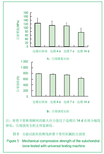
.jpg)