• 口腔组织构建 oral tissue construction • 上一篇 下一篇
成人牙根唇舌侧牙槽骨的厚度
丁继群1,方建强2,袁昌青1,陈 杰1
- 1青岛大学医学院附属医院口腔科,山东省青岛市 266003;
2杭州口腔医院正畸中心,浙江省杭州市 310006
-
收稿日期:2012-11-04修回日期:2012-11-05出版日期:2013-04-09发布日期:2013-04-09 -
通讯作者:通讯作者:袁昌青,硕士,副主任医师,主要从事口腔临床医学的研究,青岛大学医学院附属医院口腔科,山东省青岛市 266003 -
作者简介:丁继群★,女,1977年生,安徽省宣城市人,汉族,2009年温州医学院毕业,硕士,主要从事口腔临床医学的研究。
Labial and lingual alveolar bone thickness of adult tooth root
Ding Ji-qun1, Fang Jian-qiang2, Yuan Chang-qing1, Chen Jie1
- 1 Department of Stomatology, Affiliated Hospital of Qingdao University Medical College, Qingdao 266003, Shandong Province, China
2 Department of Orthodontics, Hangzhou Stomatological Hospital, Hangzhou 310006, Zhejiang Province, China
-
Received:2012-11-04Revised:2012-11-05Online:2013-04-09Published:2013-04-09 -
Contact:Yuan Chang-qing, Master, Associate chief physician, Department of Stomatology, Affiliated Hospital of Qingdao University Medical College, Qingdao 266003, Shandong Province, China chenfjq2010@163.com -
About author:Ding Ji-qun★, Master, Department of Stomatology, Affiliated Hospital of Qingdao University Medical College, Qingdao 266003, Shandong Province, China chenfjq2010@163.com
摘要:
背景:牙根在牙槽骨的位置及周围骨板厚度影响着口腔治疗,治疗过程中如果对牙齿控制不当可造成医源性并发症。以往对颌骨的研究主要针对解剖学、骨厚度或骨密度,对于牙根在牙槽骨内的空间位置及其与周围骨骼的关系,研究关注较少。 目的:建立颌骨的数字化计算机三维模型,测量牙根的唇舌侧牙槽骨厚度。 方法:选择牙列完整无明显骨骼吸收的年轻成人70例,采用牙科专用锥形束CT机进行颌面部扫描,将扫描中采集的容积信息传入计算机工作站,以及冠状位或矢状位多平面重建,获得高质量的重建图像,原始数据以DICOM格式导入计算机,并输出到整合的3D设计软件Invivo5软件进行测量。 结果与结论:重建的颌骨数字化模型可从多平面进行观察及测量,实验测得70 例患者各个牙根唇舌侧牙槽骨厚度的均值:上下前牙舌侧牙槽骨厚度大于唇侧(P < 0.05);除上前磨牙的牙颈部唇侧牙槽骨较厚外, 其他前磨牙舌侧牙槽骨厚度大于唇侧(P < 0.05);上磨牙和下颌第一磨牙唇舌侧牙槽骨厚度接近,下第二磨牙唇侧牙槽骨厚度大于舌侧(P < 0.01)。结果证实,成人不同牙位的唇舌侧牙槽骨厚度差异较大。
中图分类号:
引用本文
丁继群,方建强,袁昌青,陈 杰. 成人牙根唇舌侧牙槽骨的厚度[J]. 中国组织工程研究, doi: 10.3969/j.issn.2095-4344.2013.15.008.
Ding Ji-qun, Fang Jian-qiang, Yuan Chang-qing, Chen Jie . Labial and lingual alveolar bone thickness of adult tooth root[J]. Chinese Journal of Tissue Engineering Research, doi: 10.3969/j.issn.2095-4344.2013.15.008.
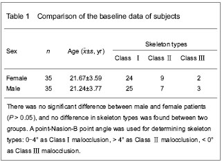
A total of 70 patients meeting the inclusion criteria were enrolled in the result analysis with no loss.
Age has a great influence on the bone and tooth root; therefore, selected subjects were all young adults. No difference was found in the baseline analysis between different genders (P > 0.05; Table 1).
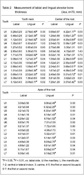
Measurement results of the labial and lingual alveolar bone thickness of the tooth root (Table 2)
| [1] Guo QY, Zhang SJ, Liu H, et al. Three-dimensional evaluation of upper anterior alveolar bone dehiscence after incisor retraction and intrusion in adult patients with bimaxillary protrusion malocclusion. J Zhejiang Univ Sci B. 2011;12(12):990-997. [2] Nahm KY, Kang JH, Moon SC, et al. Alveolar bone loss around incisors in Class I bidentoalveolar protrusion patients: a retrospective three-dimensional cone beam CT study. Dentomaxillofac Radiol. 2012;41(6): 481-488. [3] Evangelista K, Vasconcelos Kde F, Bumann A, et al. Dehiscence and fenestration in patients with Class I and Class Ⅱ Division 1 malocclusion assessed with cone-beam computed tomography. Am J Orthod Dentofacial Orthop. 2010;138(2):e1-e7.[4] Fuh LJ, Huang HL, Chen CS, et al. Variations in bone density at dental implant sites in different regions of the jawbone. J Oral Rehabil. 2010;37(5):346-351. [5] Choi JH, Park CH, Yi SW, et al. Bone density measurement in interdental areas with simulated placement of orthodontic miniscrew implants. Am J Orthod Dentofacial Orthop. 2009; 136(6):e1-e12.[6] Farnsworth D, Rossouw PE, Ceen RF, et al. Cortical bone thickness at common miniscrew implant placement sites. Am J Orthod Dentofacial Orthop. 2011;139(4): 495-503. [7] Park J, Cho HJ. Three-dimensional evaluation of interradicular spaces and cortical bone thickness for the placement and initial stability of microimplants in adults. Am J Orthod Dentofacial Orthop. 2009;136(3): e1-e12.[8] Vasak C, Watzak G, Gahleitner A, et al. Computed tomography-based evaluation of template (NobelGuide™)-guided implant positions: a prospective radiological study. Clin Oral Implants Res. 2011;22(10): 1157-1163.[9] Blok Y, Gravesteijn FA, van Ruijven LJ, et al. Micro-architecture and mineralization of the human alveolar bone obtained with microCT. Arch Oral Biol. 2012.[10] Kapila S, Conley RS, Harrell WE Jr. The current status of cone beam computed tomography imaging in orthodontics. Dentomaxillofac Radiol. 2011;40(1):24-34. [11] State Council of the People’s Republic of China. Administrative Regulations on Medical Institution. 1994-09-01.[12] Lee SL, Kim HJ, Son MK, et al. Anthropometric analysis of maxillary anterior buccal bone of Korean adults using cone-beam CT. J Adv Prosthodont. 2010;2(3):92-96.[13] Kumar V, Ludlow J, Soares Cevidanes LH, et al. In vivo comparison of conventional and cone beam CT synthesized cephalograms. Angle Orthod. 2008;78(5): 873-879.[14] Jeffrey CK, Kwong J. Image quality produced by different cone beam cephalometry tomography settings. Am J Orthod Dentofacial Orthop. 2008;133(2):317-327.[15] Suomalainen A, Vehmas T, Kortesniemi M, et al. Accuracy of linear measurements using dental cone beam and conventional multislice computed tomography. Dentomaxillofac Radiol. 2008;37(1):10-17. [16] Suomalainen A, Kiljunen T, Käser Y, et al. Dosimetry and image quality of four dental cone beam computed tomography scanners compared with multislice computed tomography scanners. Dentomaxillofac Radiol. 2009;38(6): 367-378.[17] Loubele M, Bogaerts R, Van Dijck E, et al. Comparison between effective radiation dose of CBCT and MSCT scanners for dentomaxillofacial applications. Eur J Radiol. 2009;71(3):461-468. [18] Roberts JA, Drage NA, Davies J, et al. Effective dose from cone beam CT examinations in dentistry. Br J Radiol. 2009;82(973):35-40.[19] Huang QH, Wang QB, Luo MY, et al. Xiangnan Xueyuan Xuebao. 2009;11(2):4-6.[20] Tyndall DA, Rathore S. Cone-beam CT diagnostic applications: caries, periodontal bone assessment, and endodontic applications. Dent Clin N Am. 2008;52(4):825-841.[21] Kau CH, Richmond S, Palomo JM, et al. Three-dimensional cone beam computerized tomography in orthodontics. J Orthod. 2005;32(4):282-293.[22] Ludlow JB, Ivanovic M. Comparative dosimetry of dental CBCT devices and 64-slice CT for oral and maxillofacial radiology. Oral Surg Oral Med Oral Pathol Oral Radiol Endod. 2008;106(1):106-114.[23] Zhao BD, Li NY, Zhou YG, et al. A study of rebuild of a three-dimensional anatomic model of mandibles. Huaxi Kouqiang Yixue Zazhi. 2002;20(1):21-23.[24] Januário AL, Duarte WR, Barriviera M, et al. Dimension of the facial bone wall in the anterior maxilla: a cone-beam computed tomography study. Clin Oral Implants Res. 2011;22(10):1168-1171.[25] Ghassemian M, Nowzari H, Lajolo C, et al. The thickness of facial alveolar bone overlying healthy maxillary anterior teeth. J Periodontol. 2012;83(2):187-197. [26] Henneman S, Von den Hoff JW, Maltha JC. Mechanobiology of tooth movement. Eur J Orthod. 2008; 30(3):299-306.[27] Anwar N, Fida M. Compensation for vertical dysplasia and its clinical application. Eur J Orthod. 2009;31(5):516-522.[28] Uysal T, Yagci A, Ozer T, et al. Mandibular anterior bony support and incisor crowding: Is there a relationship? Am J Orthod Dentofacial Orthop. 2012;142(5):645-653. [29] Poggio PM, Incorvati C, Velo S, et al. “Safe Zones”: A Guide for miniscrew positioning in the maxillary and mandibular Arch. Angle Orthod. 2006;76(2):191-197.[30] Kim SH, Yoon HG, Choi YS, et al. Evaluation of interdental space of the maxillary posterior area for orthodontic mini-implants with cone-beam computed tomography. Am J Orthod Dentofacial Orthop. 2009;135(5):635-641.[31] Fayed MM, Pazera P, Katsaros C. Optimal sites for orthodontic mini-implant placement assessed by cone beam computed tomography. Angle Orthod. 2010;80(5): 939-951. [32] Liou EI, Chen PH, Wang YC, et al. A computed tomographic image study on the thickness of the infrazygomatic crest of the maxilla and its clinical implications for mini screw insertion. Am J Orthod Dentofacial Orthop. 2007;131(3):352-356. [33] Lin JR, Chen S. Treatment of severe class Ⅲ with buccal shelf mini-screws. Zhonghua Kouqiang Zhengjixue Zazhi. 2010;17(3):121-126.[34] Lin JR. The new method of IZC mini-screws placement. Zhonghua Kouqiang Zhengjixue Zazhi. 2009;16(1):38-44.[35] Kwak HH, Park HD, Yoon HR, et al. Topographic anatomy of the inferior wall of the maxillary sinus in Koreans. Int J Oral Maxillofac Surg. 2004;33(4):382-388. [36] Bodner L, Brennan PA, McLeod NM. Characteristics of iatrogenic mandibular fractures associated with tooth removal: review and analysis of 189 cases. Br J Oral Maxillofac Surg. 2011;49(7):567-572. |
| [1] | 樊佳兵, 张军梅. 成年女性不同垂直骨面型下颌骨形态的测量分析[J]. 中国组织工程研究, 2021, 25(8): 1177-1183. |
| [2] | 李圆圆, 鲁颖娟, 叶玉珊, Mustafa M.M Weldali, 常少海. 构建3种不同上颌牙弓形态的有限元模型[J]. 中国组织工程研究, 2021, 25(20): 3125-3129. |
| [3] | 聂 晶, 石晓宇. 锥形束CT测量种植体支抗植入位点上颌前牙区牙槽骨厚度的性别差异[J]. 中国组织工程研究, 2021, 25(14): 2133-2136. |
| [4] | 罗诒财, 李 昊. 增强芳香烃受体表达对糖尿病模型大鼠牙槽骨缺损区愈合及炎症反应的影响[J]. 中国组织工程研究, 2021, 25(14): 2166-2171. |
| [5] | 张艺岑, 王培鑫, 刘志成. 超声引导下注射透明质酸钠与皮质类固醇治疗足底筋膜炎:疼痛、筋膜厚度及踝足功能评估[J]. 中国组织工程研究, 2021, 25(11): 1670-1674. |
| [6] | 王光平, 李明霞, 韩 雨, 徐晓梅, 徐 洁, 黄素华. 两种托槽对双颌前突患者正畸性根尖外吸收影响的比较[J]. 中国组织工程研究, 2021, 25(10): 1539-1544. |
| [7] | 许国峰, 李学斌, 唐一钒, 赵 寅, 周盛源, 陈雄生, 贾连顺. 人黄韧带细胞骨化发生过程中的自噬[J]. 中国组织工程研究, 2020, 24(8): 1174-1181. |
| [8] | 周 玉, 龙小安, 李 宁, 王 纯. 有限元法分析髌腱炎状态时的生物力学变化[J]. 中国组织工程研究, 2020, 24(8): 1280-1286. |
| [9] | 吴也可, 郜然然, 左渝陵, 余剑锋, 赵立星, 聂汶涵, 郑 涛, 艾黄萍, 严 航. 影响微种植体正畸治疗成功率的因素[J]. 中国组织工程研究, 2020, 24(4): 538-543. |
| [10] | 张 宾, 孙丽华, 张俊花, 刘童斌, 刘玉三, 崔彩云, 李 军. 上颌美学区改良盾构术与常规不翻瓣即刻种植即刻修复的短期效果比较[J]. 中国组织工程研究, 2020, 24(34): 5514-5519. |
| [11] | 覃海阔, 罗世兴. 股骨近端不同平面皮质骨厚度值、X射线灰度值与女性髋部脆性骨折的相关性[J]. 中国组织工程研究, 2020, 24(18): 2867-2872. |
| [12] | 程余婷, 伍 超, 黄晓林, 李 芳, 石前会, 周 倩, 洪 伟, 王 永, 廖 健. 低剂量唑来膦酸对去势拔牙大鼠破骨及成骨细胞的影响[J]. 中国组织工程研究, 2020, 24(17): 2686-2693. |
| [13] | 张钰晶, 彭于治, 刘宝珍, 井 芳. 锥形束CT评价仙灵骨葆胶囊治疗绝经后女性牙周炎患者牙槽骨骨量的变化[J]. 中国组织工程研究, 2020, 24(17): 2694-2699. |
| [14] | 陈周艳, 周 容, 何 淞, 吴定丹, 吴 稀, 白 蕊, 黄 跃. 矩形附件厚度与位置变化对矫正尖牙扭转的影响#br#[J]. 中国组织工程研究, 2020, 24(16): 2513-2519. |
| [15] | 宋旭东, 何云武, 李勇霖, 陈 静, 胡君兰. 超声引导下椎旁神经阻滞治疗带状疱疹相关疼痛的Meta分析[J]. 中国组织工程研究, 2020, 24(11): 1797-1804. |
Orthodontic braces are used in orthodontics to align and move teeth to the desired positions
with regard to a person’s bite. Alveolar bone defects are very common prior to orthodontic treatment[2]. During tooth movement, three-dimensional cone-beam CT images are important to avoid iatrogenic periodontal support loss of anterior teeth, especially the lingual bone
plate of lower incisors[3].
A observational experiment.
The experiment was conducted in the Department of Orthodontics, Hangzhou Stomatological Hospital, China from October 2010 to May 2012.
Seventy adult patients with malocclusion from the Department of Orthodontics, Hangzhou Stomatological Hospital were enrolled, including 35 females and 35 males.
Inclusive criteria
Patients aged 18-25 years had complete dentition in the study area (excluding third molars) confirmed via clinical and X-ray examination, and presented no significant bone absorption (Figure 1).
.jpg)
Exclusion criteria
Patients with dental disease, apical shadow, jaw cysts or tumors were excluded. Patients who took drugs affecting bone metabolism within 6 months or those with hyperthyroidism and diabetes mellitus were excluded.
.jpg)
Cone-beam CT scanning and establishing a digital three-dimensional model of the adult jaw
Dental cone beam CT machine was adopted for maxillofacial scanning. Patients sat to place their heads on the scan head frame, and the infraorbital line was parallel to the ground.
.jpg)
Measuring the labial and lingual alveolar bone thickness of the tooth root and relevant parameters
In vivo 5 image analysis software was employed for three-dimensional image processing. Reconstruction images were cut according to the reference line and angle, and alveolar bone thickness was measured according to Lee’s method[12], as shown in Figure 3.
(1) Thickness of the alveolar bone plates Measurement method.
(2) Labial alveolar bone thickness of the dental cervix: the distance from the most former (outside) point to the most-back (inner) point along the cervix vertical line.
(3) Lingual alveolar bone thickness of the dental cervix: the distance from the most former (outside) point to the most-back (inner) point along the cervix vertical line.
(4) Labial alveolar bone thickness of the middle of the root: the distance from the most former (outside) point to the most-back (inner) point along the central vertical line of the root.
(5) Lingual alveolar bone thickness of the middle of the root: the distance from the most former (outside) point to the most-back (inner) point along the central vertical line of the root.
(6) Labial alveolar bone thickness of the root apex: the distance from the most former (outside) point to the most-back (inner) point along the apical vertical line.
.jpg)
Measurement of parameters is shown in Figures 4-6.
.jpg)
.jpg)
Main outcome measurements
Labial and lingual alveolar bone thickness of the dental cervix; labial and lingual alveolar bone thickness of the middle of root; labial and lingual alveolar bone thickness of the root apex.
Measurement data were expressed as mean±SD, and data analysis was performed using SPSS 17.0 statistical software. Mean differences between groups were compared using paired t-test, and a value of P < 0.05 was considered significant.
Reliability of cone-beam CT
Conventional anatomy is mainly focused on the maxilla general form, and there is lack of therapeutic guidance for clinical orthodontics and alveolar surgery. X-ray imaging is an important means of research. However, because of the serious overlap of the two-dimensional images, such as conventional cephalometric films, low resolution, and high radiation, ordinary CT cannot clearly display the fine structure of local bone tissues and any tooth points, leading to physician’s difficulty in determining the relationship between the teeth and surrounding structures[13-14]. Cone-beam CT provides the best choice[9, 15-18]. Domestic scholars have reported cadaveric anatomic studies focusing on the alveolar height[19], and CT studies of bone mineral density[4-5] and cortical bone thickness[6-7]. But, there is still a lack of data regarding the spatial location of the root within the alveolar bone and its relationship with the surrounding bone, which is worthy of attention.
Cone-beam CT is flexible for three-dimensional imaging, minimizes the radiation dose to obtain high-resolution three-dimensional scanning results, provides a smooth soft tissue image, displays the hard tissue and bone structure detail and contrast to the maximal extent, and clearly show the tooth, root apex, alveolar bone, maxillary sinus and mandibular canal, providing a full range of diagnostic information for dental, periodontal, implanting, orthodontic, maxillofacial surgery, and repair treatment[20]. Cone-beam CT has high resolution[8], clear image quality, shorter scan time, and low ray dose[21-22] to supply low-radiation-dose and high-definition images for dentistry, greatly improving the diagnostic accuracy and cure rates of oral diseases, with a wide range of applications.
Gross observation showed non-equivalent thickness of the labial/buccal and palatal/lingual bone plates of the maxillary tooth socket. The present study found that labial bone plate thickness of the upper teeth showed a gradual thickening from front to back, while the lingual bone plate thickness gradually decreased at the middle and apex of the tooth root, especially in the cervical part of the canine and first premolar at the lingual side and in the root middle of the first and second premolars at lingual side, showing a narrowing phenomenon. Labial/lingual bone plates of the lower teeth gradually thickened from front to back, showing a gradual thickening tendency from the cervical to the apical side. This measurement results were consistent with those of Zhao et al [23].
The scope and significance of orthodontic tooth movement
No definitive conclusion has been defined for the scope of orthodontic tooth movement. Concept of dentition boundaries gives a rough reference for orthodontic tooth movement, but,with regard to disputes, there is no quantitative standard. Generally, thickness of the alveolar bone around the root is considered as the boundary of tooth movement that refers to the dense cortical bone plate at the same level. Apical tooth movement should not exceed this boundary. Lingual alveolar bone plate of the lower incisors was significantly thicker than the labial, confirmed by CT measurements[2]. The lingual alveolar bone plate of the upper incisors was thicker than the labial thicker, indicating that appropriate incisor adduction can improve the prognathism face commonly seen in Chinese people[1]. It is often necessary to control the root of the maxillary anterior teeth facing to the tongue, but the key is to grasp the adductor limit and anatomy restrictions for the alveolar bone cannot be ignored. Excessive adduction can result in the contact between the root and dense cortical bone plate to increase the risks. The labial bone plate of the neck of the anterior teeth, especially the lower incisors, was very thinner, and even presented no bone coverage; and a thinner bone plate was also found at the labial side of the middle of the root. During the process of tooth movement, biological alterations in the alveolar bone are mainly focused on bone resorption, and bone hyperplasia is very limited[26], prompting that we will attach importance to the labial inclination of the anterior teeth. Excessive labial inclination can result in gingival recession, washboard-shaped root and even fenestration of the tooth apex via the alveolar bone plate. In the present study, the alveolar bone thickness of the maxillary first premolar at the root neck was (1.35±0.67) mm, and the labial bone plate thickness of the first molar at the root neck was (2.85±0.80) mm. During rapid palatal expansion, it should pay attention to preventing against dehiscence and gingival recession in elderly patients. In clinical orthodontic tooth movement, in particular, in patients accompanied with a certain degree of bone abnormalities, conceal orthodontic treatment often requires a certain compensation for the location and angle of the teeth[27]. It should pay attention to the relationship between the root and the alveolar bone, especially in high-risk groups with poor anatomical structures and periodontal status. When necessary, CT assessment can be used to prevent against iatrogenic problems that affects tissue health and treatment stability[28].
Labial area of the maxillary second premolar and first molar is the most common region for orthodontic anchorage screw implantation[29-31]. In the present study, the region with the thickest buccal bone plate was the appropriate site for orthodontic anchorage screw implantation. The labial bone plate at the root apex of the maxillary second molar was thinner than that of the maxillary first molar, which is inconsistent with the result of Liou et al [32]. It is likely associated with the observed region in the present study only focusing on the apical parts rather than the zygomatic crest area. Under the relevant tips from Lin et al [33], orthodontic anchorage implantation outside of the tooth root should be carefully performed at the maxillary first and second molar area to move the upper dentition as a whole. However, the number of cases may be low for this method. The maxillary palatal space is generally higher than the labial, providing more space for implantation. The second premolar region is the thickest in the palatal parts of the maxillary posterior teeth, suggesting patients who are difficult in buccal application can select this region for implantation. The buccal parts of the mandibular molars, especially the buccal bone plate of the mandibular second molar, significantly become thicker, rendered as a platform, which is called as “buccal shelf area”[34]. In fact, the corresponding anatomical structure is the external oblique, providing sufficient implant space, which is an ideal area for anchorage screw implantation.
Root fracture is the most common complication of tooth extraction surgery, which is mainly caused by no fully understanding about anatomical structures surrounding the extracted teeth and use of violence. Extraction force should be applied directing to the small bone resistance and avoid violence. This study showed that the lingual bone plates of the root of the anterior teeth and premolars were generally thicker than the labial; the palatal bone plate of the maxillary molar region was significantly thickened, indicating that extraction force should be applied towards the labial and buccal sides, to avoid bone resistance. Meanwhile, extraction force should be soft to avoid root fracture and even damage to the maxillary sinus, as the root of the maxillary molar was approaching the maxillary sinus[35]. Because of the obvious thickening of the buccal bone plate in the mandibular molar region, extraction force should lean to the lingual part with small bone resistance, which is conducive to tooth dislocation, and not easy to cause root fracture or jaw fracture[36].
1.中国关于牙根及周围牙槽骨的研究关注较少,均为采用口腔专用的锥形束CT扫描。国外有锥形束CT关于骨密度、前牙牙槽骨厚度的研究,仅有的牙槽骨厚度研究均为上颌前牙区域,实验利用锥形束CT扫描获得中国人上下颌骨全部牙齿的唇舌侧牙槽骨厚度数据。 2. 结果证实,成人不同牙位的唇舌侧牙槽骨厚度差异较大。
1文章构思特点 颌骨和牙齿是口腔医学的基础,对颌骨的局部或系统解剖及牙齿相关结构已进行大量研究,对颌骨及牙齿的治疗、手术有重要的指导意义,但由于牙根被牙槽窝周围的固有牙槽骨包绕,对于牙根在牙槽骨的位置特别是周围牙槽骨厚度,常规手段判断困难,本实验拟采用锥形束CT探讨牙根唇舌侧牙槽骨厚度及其对口腔个性化治疗的意义。 2本文创新点: ①数据库为CNKI和 Medline,检索时间为截止2012年,检索关键词设定为牙槽骨(alveolar bone)牙根(root of tooth)、厚度( thickness)、锥形束CT(Cone-beam CT)最终检索认定本文(实验)具先进性。 ②创新性说明:中国关于牙根及周围牙槽骨的研究关注较少,朱胜吉等用CT研究上颌后牙区解剖特点,姜若萍等用侧位片研究成年人前牙区牙槽宽度及根尖位置,没有采用口腔专用的锥形束CT扫描。经检索发现国外有锥形束CT关于骨密度、前牙牙槽骨厚度的研究,仅有的牙槽骨厚度研究均为上颌前牙区域,本研究利用锥形束CT扫描获得中国人上下颌骨全部牙齿的唇舌侧牙槽骨厚度数据。
| 阅读次数 | ||||||
|
全文 |
|
|||||
|
摘要 |
|
|||||