中国组织工程研究 ›› 2013, Vol. 17 ›› Issue (1): 62-67.doi: 10.3969/j.issn.2095-4344.2013.01.010

• 骨髓干细胞 bone marrow stem cells • 上一篇    下一篇

兔骨髓间充质干细胞分离培养后的活力检测

孔根现12,蒋知新12,沙 杭2,张清华12   

  1. 1南方医科大学研究生学院,广东省广州市 510515
    2解放军第三○五医院老年病中心,北京市 100017
  • 收稿日期:2012-05-17 修回日期:2012-07-05 出版日期:2013-01-01 发布日期:2013-01-01
  • 通讯作者: 张清华,主任医师,南方医科大学研究生学院,广东省广州市 510515;解放军第三○五医院老年病中心,北京市100017
  • 作者简介:孔根现★,男,1986年生,河南省开封市尉氏县人,汉族,南方医科大学在读硕士,主要从事老年心血管病的研究。312973139@qq.com
  • 基金资助:

    国家自然科学基金资助(30971235),课题名称:骨髓干细胞动员与移植稳定修复易损斑块的实验研究。

Isolation, culture and cell viability testing of rabbit bone marrow mesenchymal stem cells

Kong Gen-xian1,2, Jiang Zhi-xin1,2, Sha Hang2, Zhang Qing-hua1,2   

  1. 1 Postgraduate School, Southern Medical University, Guangzhou 510515, Guangdong Province, China
    2 Geriatric Center, the 305 Hospital of Chinese PLA, Beijing 100017, China
  • Received:2012-05-17 Revised:2012-07-05 Online:2013-01-01 Published:2013-01-01
  • Contact: Zhang Qing-hua, Chief physician, Postgraduate School, Southern Medical University, Guangzhou 510515, Guangdong Province, China; Geriatric Center, the 305 Hospital of Chinese PLA, Beijing 100017, China
  • About author:Kong Gen-xian★, Studying for master’s degree, Postgraduate School, Southern Medical University, Guangzhou 510515, Guangdong Province, China; Geriatric Center, the 305 Hospital of Chinese PLA, Beijing 100017, China 312973139@qq.com
  • Supported by:

    the National Natural Science Foundation of China, No. 30971235*

摘要:

背景:目前分离纯化骨髓间充质干细胞多采用单一的方法,对扩增培养过程中细胞活力的变化没有较为系统而全面的评估。

目的:探索体外分离培养和纯化兔骨髓间充质干细胞的方法,并对细胞的活力进行检测。

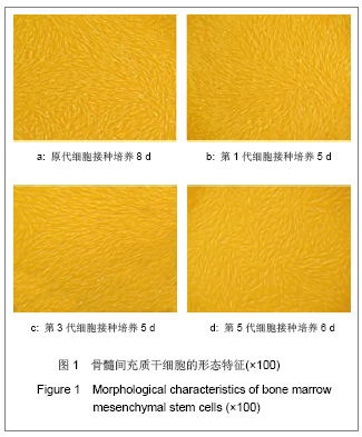
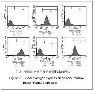
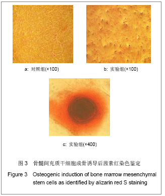
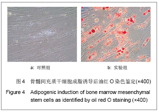
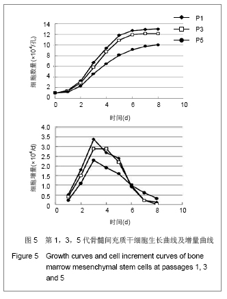
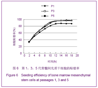
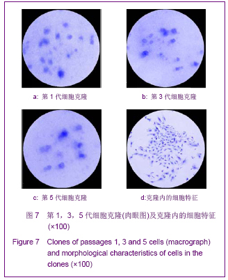
方法:采用密度梯度离心联合贴壁筛选的方法分离纯化兔骨髓间充质干细胞,从形态学、细胞表型和成骨成脂能力方面进行鉴定;并从细胞生长曲线、贴壁率和克隆形成率3个方面评估细胞活力。

结果与结论:原代细胞多呈梭形和圆形,48 h后大部分细胞贴壁,8-10 d细胞融合达80%-90%,传代周期为3-5 d;流式检测显示CD14、CD34和CD45表达阴性,CD29、CD44和CD90表达阳性;成骨诱导茜素红染色可见明显的钙结节,成脂诱导油红O染色可见大量脂肪细胞;细胞生长曲线、贴壁率及克隆形成率的结果表明第1代和第3代细胞的活力优于第5代细胞。结果说明密度梯度离心联合贴壁筛选的方法能够有效的分离纯化骨髓间充质干细胞,并能较好的保持其生物功能,在扩增传代过程中,细胞活力会有不同程度的下降。

关键词: 干细胞, 骨髓干细胞, 骨髓间充质干细胞, 分离纯化, 鉴定, 生长曲线, 贴壁率, 克隆形成率, 国家自然科学基金, 干细胞图片文章

Abstract:

BACKGROUND: Most of recent studies on isolation and purification of bone marrow mesenchymal stem cells adopt single methods, and there is no systematic and comprehensive evaluation on cell viabilities during amplification.
OBJECTIVE: To establish an effective method of purifying rabbit bone marrow mesenchymal stem cells and to test the cell viabilities.   
METHODS: Rabbit bone marrow mesenchymal stem cells were isolated by density gradient centrifugation and adherent cultivation methods. Rabbit bone marrow mesenchymal stem cells were identified from the morphology, cell phenotype and osteogenic and adipogenic capacity. Cell viabilities were evaluated using growth curve, seeding efficiency and colony-forming efficiency.
RESULTS AND CONCLUSION: The primary cells were spindle-shaped and circular. Most of the cells attached to the dish after seeding for 48 hours. After 8 or 10 days, cells reached 80%-90% confluency and the subculture cycle was 3 to 5 days. Flow cytometry demonstrated that cells were negative for CD14, CD34 and CD45, but they were positive for CD29, CD44 and CD90. Calcium nodules were observed by alizarin red S staining. A large amount of adipose-derived stem cells were observed by oil red O staining. Cell viability of passages 1 and 3 was stronger than that of passage 5. These findings suggest that density gradient centrifugation combined with adherent cultivation can be used to effectively isolate and purify bone marrow mesenchymal stem cells and well maintain the proliferation and differentiation potential of bone marrow mesenchymal stem cells. Cell viability will be decreased to different extents during cell proliferation.
Key Words: stem cells; bone marrow-derived stem cells; bone marrow mesenchymal stem cells; isolation and purification; identification; growth curve; adherence rate; clone formation rate; National Natural Science Foundation of China; stem cell photographs-containing paper

Key words: stem cells, bone marrow-derived stem cells, bone marrow mesenchymal stem cells, isolation and purification, identification, growth curve, adherence rate, clone formation rate, National Natural Science Foundation of China, stem cell photographs-containing paper

中图分类号: